The DME Perfusion Paradox
In the clinical management of diabetic macular edema (DME), the advent of anti-VEGF therapy has revolutionized our ability to achieve anatomical and functional success. We routinely observe significant reductions in central subfield thickness (CST) and the robust resolution of intraretinal and subretinal fluid. However, a “perfusion paradox” persists: despite the effective “drying” of the macula, the underlying retinal nonperfusion index (NPI)—a critical marker of disease severity and a harbinger of progression—often remains recalcitrant to therapy.
The KINGFISHER trial provided a rigorous framework to examine this disconnect. While the primary study established brolucizumab’s anatomical superiority over aflibercept, a subsequent post hoc analysis was undertaken to determine if this enhanced fluid resolution translated into measurable microvascular reperfusion. This analysis compared longitudinal changes in NPI in eyes treated with intensive monthly brolucizumab versus aflibercept over a 52-week period.
Study Foundation and Methodology
This investigation was a post hoc analysis of the multicenter, masked KINGFISHER trial. The initial cohort consisted of 517 participants, from which 164 eyes had available baseline ultrawidefield fluorescein angiography (UWF-FA). To ensure precise quantification of nonperfusion, researchers excluded 43 cases with prior laser photocoagulation scars and 30 cases with inadequate image quality, resulting in a final cohort of 91 eyes (59 brolucizumab, 32 aflibercept).
The baseline characteristics and retinopathy distribution of the cohort are summarized below:
| Characteristic | Total Cohort (n=91) | Brolucizumab (n=59) | Aflibercept (n=32) | p-Value |
| Age (years) | 59.6 ± 10.2 | 58.9 ± 11.5 | 61 ± 7.2 | 0.273 |
| Gender (Male, %) | 56 (61.5%) | 35 (59.3%) | 21 (65.6%) | 0.555 |
| DM Duration (years) | 11.4 ± 17.8 | 13.4 ± 20.4 | 7.8 ± 10.8 | 0.090 |
| BCVA (ETDRS letters) | 59.9 ± 9.8 | 59.5 ± 10.0 | 60.5 ± 9.6 | 0.646 |
| CST (µm) | 534.1 ± 157.7 | 547.6 ± 165.8 | 509.5 ± 140.9 | 0.273 |
| DRSS Distribution (n) | 0.723 | |||
| Level 35 / 43 | 14 / 16 | 9 / 10 | 5 / 6 | |
| Level 47 / 53 / 61 | 32 / 12 / 13 | 20 / 9 / 9 | 12 / 3 / 4 |
UWF-FA images were graded by the Doheny Image Reading Center using Optos Advance software. Three regions of interest (ROI) were analyzed: the Total Analyzed Retina (TAR), the Posterior Pole (a 15mm foveal circle), and the Peripheral Zone (TAR minus the posterior pole).
Key Findings: The “No Change” Reality
The 52-week data revealed a sobering reality: measurable retinal nonperfusion remained essentially stable despite intensive monthly anti-VEGF therapy. There was no statistically significant change in NPI across any retinal zone for either drug.
| Retinal Zone | Drug | Baseline NPI (%) | Week 52 NPI (%) |
| Posterior Pole | Brolucizumab | 6.1 ± 8.3 | 6.3 ± 10.5 |
| Aflibercept | 3.3 ± 4.2 | 3.6 ± 6.0 | |
| Peripheral Zone | Brolucizumab | 18.2 ± 17.4 | 18.0 ± 18.6 |
| Aflibercept | 12.4 ± 12.7 | 13.0 ± 12.2 | |
| Pan-retinal (TAR) | Brolucizumab | 15.2 ± 14.2 | 15.1 ± 15.9 |
| Aflibercept | 10.3 ± 9.9 | 10.7 ± 9.8 |
Linear mixed-effects modeling confirmed these longitudinal trends:
- Stability over Reperfusion: Neither brolucizumab nor aflibercept induced significant reperfusion or significant worsening.
- Non-Significant Interaction: The visit-by-treatment interaction was non-significant (p > 0.05 for all zones; p=0.91 for pan-retinal), indicating neither agent was superior in altering the ischemic trajectory.
- Baseline Imbalance: Brolucizumab eyes exhibited numerically higher NPI at baseline, representing a numerical imbalance rather than a treatment-induced divergence.
Deep Dive: Anatomical Success vs. Ischemic Stability
The primary KINGFISHER trial demonstrated that brolucizumab’s high molar concentration and enhanced bioavailability resulted in superior fluid drying. However, this post hoc analysis highlights that “potency” in fluid resolution does not equate to “reversibility” of ischemia.
The data supports the theory that established retinal nonperfusion may involve irreversible structural changes, such as endothelial cell proliferation and permanent vascular occlusion, rather than merely transient, reversible events like vascular leukostasis (leukocyte plugs). While neutralizing VEGF may resolve the leukostatic component, it appears insufficient to address chronic capillary death.
These findings align with and expand upon evidence from other major trials:
- RISE/RIDE and VISTA: Suggested that anti-VEGF slows the progression of NPI compared to sham, but does not provide true reperfusion.
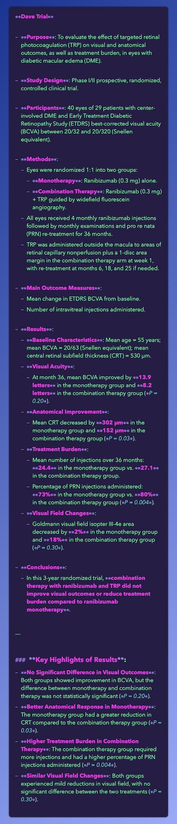
- DAVE and CLARITY Substudy: Reinforced the observation that even with intensive therapy, existing areas of nonperfusion remain largely static.
- RECOVERY and PERMEATE: Observed that NPI stability is highly dependent on dosing consistency, noting potential worsening when moving from monthly to extended intervals.
Clinical Implications for Retina Specialists
The Role of VEGF and “Clinical Pearls” For the clinician, the primary takeaway is that anatomical “normalization” on OCT does not imply microvascular recovery on UWF-FA. While VEGF is a potent driver of permeability, its inhibition is not a “magic bullet” for reopening occluded capillaries. Clinicians should manage expectations: even a perfectly dry retina may harbor significant, unchanging ischemia.
Non-VEGF Pathways The persistence of NPI suggests that the ischemic cascade is maintained by pro-inflammatory cytokines and growth factors beyond the VEGF family. Future therapeutic efforts may require multi-pathway inhibition to address the irreversible components of diabetic microangiopathy and promote functional “outsprouting” of new vessels.
Dosing Regimens The stability observed in KINGFISHER occurred under a strict monthly regimen. Academic speculation remains regarding whether the superior potency of brolucizumab could maintain this stability at extended intervals—such as quarterly dosing—or if continuous, high-frequency inhibition is the only way to prevent ischemic progression.
Study Limitations and Future Directions
- Methodological Constraints: As a post hoc analysis, the study is susceptible to selection bias and lacks an untreated/sham control group to establish definitive causality for the observed stability.
- Sample and Scope: The relatively small sample size (n=91) and the 52-week duration may lack the power to detect subtle, long-term shifts in peripheral perfusion.
- Grading Challenges: Manual delineation of NPI remains subjective and time-intensive compared to emerging automated algorithms.
Conclusion: Final Summary
measurable retinal nonperfusion remains largely unchanged over 52 weeks in DME patients receiving monthly anti-VEGF therapy. While brolucizumab provides superior anatomical drying compared to aflibercept, neither agent facilitates significant microvascular reperfusion. The stability of the nonperfusion index suggests that while we can mitigate the effects of vascular leakage, the underlying structural ischemia represents a predominantly irreversible aspect of diabetic eye disease.
Bottom Line: Clinicians should treat for fluid but recognize that ischemic stability—not reversal—is the realistic benchmark for current anti-VEGF protocols. Further longitudinal studies into multi-pathway inhibition are required to move the needle on retinal reperfusion.

Alhelaly, Mai, et al. “Comparison of Retinal Nonperfusion in Diabetic Macular Edema Treated with Brolucizumab Versus Aflibercept: A KINGFISHER Trial Analysis.” Am. J. Ophthalmol., vol. S0002-9394, no. 26, 23 July 2026, pp. 00419-8, doi:10.1016/j.ajo.2026.07.041.