
Introduction: Defining a Non-Pathognomonic Sign
For decades, the presence of curvilinear peripheral streaks—known eponymously as Schlaegel lines (SL)—has served as a subtle yet significant clinical clue in the evaluation of chorioretinal disorders. First described in 1981 by James A. Fountain and Theodore F. Schlaegel Jr. as the “fourth cardinal sign” of presumed ocular histoplasmosis syndrome (POHS), these lesions were originally thought to be unique to Histoplasma-endemic regions like the Ohio River Valley. However, our understanding has evolved. Today, SL are recognized as a distinctive, though non-pathognomonic, ophthalmoscopic sequela of various inflammatory and, surprisingly, inherited retinal diseases (IRDs).
For the retina specialist, recognizing SL is not merely an exercise in naming a rare finding; it is a critical step in identifying underlying systemic or ocular pathologies and assessing the risk for vision-threatening complications like macular neovascularization (MNV).
The Clinical Profile: Who Is at Risk?
A comprehensive analysis of current literature reveals a clear demographic predilection. SL predominantly affect young to middle-aged women, with a female-to-male ratio of approximately 2.3:1. The mean age at presentation is roughly 26.7 years, though the range is broad (9 to 66 years).
Perhaps the most consistent clinical association is refractive error. Approximately 68% of patients with SL exhibit myopic refraction, with a mean spherical equivalent of -6.7 diopters. This strong link to moderate-to-high myopia suggests that equatorial stretching and axial elongation may play a role in the pathogenesis or progression of these lesions.
Etiological Spectrum: From Uveitis to IRDs
While originally tethered to POHS, the etiological landscape of SL is now dominated by inflammatory chorioretinal disorders, which account for 83% of documented cases.
- Multifocal Choroiditis and Punctate Inner Choroidopathy (MFCPU/PIC): Currently the leading cause, accounting for 43.4% of cases. In these patients, SL appear as linear clusters of atrophic spots that may represent the coalescence of focal inflammatory infiltrates.
- Presumed Ocular Histoplasmosis Syndrome (POHS): The second most common etiology at 37.3%. Here, SL are often viewed as a linear aggregation of “histo spots”.
- Inherited Retinal Diseases (IRDs): Surprisingly, 17% of SL cases occur in the context of IRDs, most notably Pseudoxanthoma elasticum (PXE) and Stargardt disease. In PXE, severe disruption of the Bruch membrane (BM) may trigger a secondary inflammatory response resembling PIC, leading to the formation of SL.
- Rare Associations: Isolated cases have been linked to Vogt-Koyanagi-Harada (VKH) syndrome and Acute Posterior Multifocal Placoid Pigment Epitheliopathy (APMPPE).
Pathophysiology: The Watershed and Pressure Theories
The mechanism behind the characteristic linear arrangement remains a subject of debate, though two primary theories prevail:
- The Vascular Watershed Theory: Originally proposed by Fountain, this theory suggests that the equatorial choroidal vasculature—supplied by a bidirectional system of posterior and recurrent anterior ciliary arteries—creates a watershed zone. This anatomical configuration may facilitate the preferential deposition of immunogenic material or antigen-antibody complexes, triggering localized inflammation.
- The Tangential Pressure Hypothesis: Some researchers propose that focal lymphocytic infiltrations exert tangential pressure on the retinal pigment epithelium (RPE), causing lesions to expand laterally and eventually coalesce into curvilinear streaks.
Multimodal Imaging: Characterizing the Streaks
Modern imaging is indispensable for the diagnosis and longitudinal monitoring of SL.
- Fundus Examination and Photography: SL appear as punched-out chorioretinal atrophic spots or streaks, typically located in the mid-to-far periphery. They are oriented parallel to the ora serrata and can extend from 15° to a full 360° circumference.
- Fundus Autofluorescence (FAF): Inactive SL demonstrate hypo-FAF due to RPE loss, often surrounded by a hyper-FAF margin that may indicate evolving inflammation.
- Optical Coherence Tomography (OCT):
- Inactive lesions: Show inhomogeneous disruption of the RPE-BM-choriocapillaris complex.
- Active lesions: May display hump-like hyperreflective RPE elevations with associated choroidal hypertransmission.
- En Face OCT: SL appear as hyporeflective curvilinear lesions in the outer retinal slab and show flow deficits on OCT Angiography (OCTA) at the level of the choriocapillaris.
- Fluorescein Angiography (FA) and ICGA: FA typically shows RPE window defects (early hyperfluorescence), while ICGA consistently demonstrates hypocyanescence across all phases, reflecting permanent choroidal scarring.
The Major Complication: Macular Neovascularization (MNV)
The clinical significance of SL is heightened by their association with Type 2 MNV, which develops in 44% of eyes. This vision-threatening complication is most prevalent in POHS (59%) and MFCPU/PIC (47%), but can also occur in IRD-associated cases (17%). Interestingly, patients with SL may exhibit multiple MNV lesions (up to 6 in some reports), necessitating vigilant long-term follow-up.
Differential Diagnosis: Avoiding Mimics
Several conditions can mimic SL, and differentiation is critical for management:
- Peripheral Lacquer Cracks: These are mechanical fissures of the BM due to pathologic myopia. Unlike the atrophic clusters of SL, lacquer cracks are linear breaks without the same inflammatory morphology.
- West Nile Virus (WNV) Chorioretinitis: Presents with linear clusters, but they follow a radial, neurotropic orientation tracking the retinal nerve fiber layer (RNFL), unlike the circumferential pattern of SL.
- Verhoeff Lines: These are pigmented ridges marking the boundaries of previous choroidal detachments. They are typically concave with respect to the fovea, whereas SL are convex.
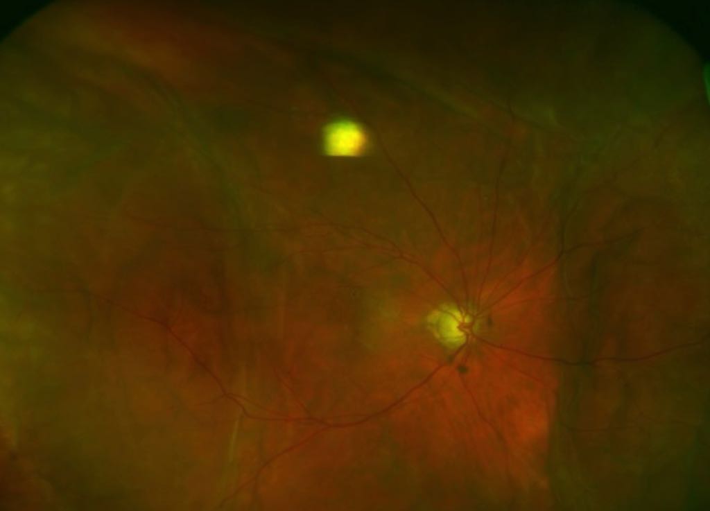
4. Lattice Degeneration: Affects the neurosensory retina rather than the chorioretinal interface and is associated with vitreoretinal adhesion and hyalinized vessels.
Conclusion: Clinical Pearls for the Retina Clinic
Schlaegel lines are more than just peripheral curiosities; they are markers of significant chorioretinal pathology. The high prevalence of associated MNV makes ultra-widefield (UWF) imaging an essential tool for early detection and monitoring. When SL are identified, the specialist must:
- Consider an underlying inflammatory etiology (MFCPU/PIC or POHS).
- Rule out IRDs like PXE if systemic signs are present.
- Distinguish from mechanical (lacquer cracks) or infectious (West Nile) mimics using orientation and multimodal patterns.
- Monitor closely for the development of macular complications.
As we move forward, longitudinal studies are needed to determine if the expansion or repigmentation of SL can serve as reliable biomarkers for disease activity and therapeutic response.

Reference Paolo Forte, Alessandro Feo, et al. “Schlaegel lines: History, etiology, multimodal imaging, and differential diagnosis of curvilinear streaks in chorioretinal disorders.” Survey of Ophthalmology. Available online May 2026.


