
1. Introduction: The Collapse of the Binary Paradigm
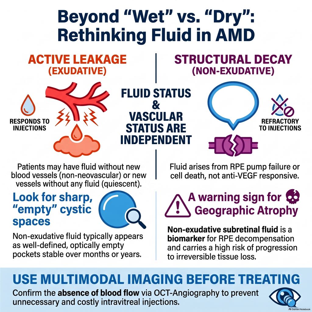
By 2026, the long-standing clinical management of age-related macular degeneration (AMD) has undergone a fundamental shift. For decades, we relied on a simplistic binary framework: “dry” (non-exudative) versus “wet” (exudative). Within that traditional paradigm, detecting any fluid on structural optical coherence tomography (OCT) was considered a pathognomonic sign of neovascular activity, mandating immediate and chronic anti-vascular endothelial growth factor (anti-VEGF) therapy.
This “fluid equals treatment” dogma has collapsed under the weight of high-resolution multimodal imaging (MMI). We now recognize non-exudative fluid—also referred to as non-vasogenic cystoid maculopathy or transudative hyporeflective spaces—as a distinct clinical phenotype. These spaces arise from degenerative, mechanical, or compensatory failures rather than active macular neovascularization (MNV). Distinguishing these entities is vital to prevent over-treatment, reduce patient burden, and accurately identify eyes at high risk for geographic atrophy (GA) progression.
2. The New Clinical Framework: The 2×2 Matrix
Our current standard of care requires the independence of “Vascular Status” (Neovascular vs. Non-neovascular) and “Fluid Status” (Exudative vs. Non-exudative). These represent two separate axes of disease progression.
| Neovascular (MNV Present) | Non-Neovascular (MNV Absent) | |
| Exudative (Fluid Present) | Neovascular Exudative: Standard “wet” AMD; active leakage requiring anti-VEGF. | Non-Neovascular Exudative: Fluid arising from a non-MNV-mediated breakdown of the outer blood-retinal barrier (oBRB) or retinal capillary integrity (e.g., in end-stage atrophic retina). |
| Non-Exudative (No Fluid / Non-exudative Fluid) | Neovascular Non-exudative: “Quiescent” or subclinical MNV; high risk (20-40%) for exudative conversion. | Non-Neovascular Non-exudative: Standard “dry” AMD or degenerative/transudative non-vasogenic spaces. |
Quiescent MNV is the conceptually inverse entity of non-exudative fluid: a neovascular network detectable on OCT angiography (OCT-A) that lacks associated fluid and typically requires observation rather than injection.
3. Multimodal Imaging (MMI) as the Diagnostic Bedrock
Structural OCT is our most sensitive tool for detection, but it is insufficient for determining the pathophysiology of fluid. Confirmation of a non-exudative state relies on negative diagnostic criteria across MMI modalities:
- Fluorescein Angiography (FA) and ICGA: Non-exudative fluid exhibits an absence of progressive, ill-defined leakage or pooling. While FA may show “window defects” in GA, it lacks the active staining of a neovascular source.
- OCT Angiography (OCT-A): This provides depth-resolved visualization to confirm the absence of an abnormal flow signal in the outer retina or sub-RPE space at the fluid’s site.
Technical Perspective: Addressing False-Negatives
Clinicians must remain vigilant regarding false-negative OCT-A results that may mask active MNV, particularly:
- Segmentation Errors: Misplacement of slabs in distorted anatomy.
- Media Opacities: Signal attenuation from cataracts or vitreous haze.
- Shadowing: Signal loss beneath pigment epithelial detachments (PED) or subretinal hyperreflective material (SHRM).
- Slow-Flow Lesions: Type 1 MNV or small, early Type 3 MNV that may fall below the flow-detection threshold.
4. Decoding OCT Structural Biomarkers: Exudative vs. Non-Exudative
Differentiating true exudation from transudative spaces requires meticulous analysis of internal reflectivity, shape, and temporal stability.
| Feature | Exudative Neovascular Fluid | Non-Exudative Fluid |
| Histological Composition | Protein-rich exudate; contains fibrin, blood cells, and inflammatory mediators. | Acellular, protein-poor transudate or degenerative space containing tissue debris. |
| Internal Reflectivity | Granular or faintly hyperreflective signal. | Homogeneously hyporeflective; optically “empty” lumen. |
| Morphological Shape | Rounded, poorly delineated contours; interacts dynamically with tissue. | Square-edged, angular, or sharply demarcated contours. |
| Retinal Layer | Often outer retina or associated with focal outer retinal disruption. | Inner Nuclear Layer (INL) for pseudocysts; Henle fiber layer in nascent GA. |
| Temporal Behavior | Dynamic; rapid onset and short-term fluctuation. | Highly stable; configuration persists for months/years. |
| Anti-VEGF Response | Responsive; fluid volume typically decreases or resolves. | Refractory; fluid is unaffected by injections. |
5. Pathophysiology of Non-Exudative Intraretinal Fluid (IRF)
Non-exudative IRF is driven by three distinct mechanisms unrelated to vascular permeability:
- Degenerative Processes: In nascent GA, hyporeflective spaces often follow the course of the Henle fiber layer. As atrophy establishes, Müller cells (the primary glial support) undergo apoptosis, leading to retinal cavitation. These “Degenerative Pseudocysts” are most numerous in the inner nuclear layer (INL) and represent tissue loss rather than true fluid accumulation.
- “Burnt-out” Contraction: Chronic fibrotic scars, though no longer leaking, contain densely collagenized matrix that may contract over time. This architectural distortion exerts tractional forces on the overlying retina, disrupting Müller cell columns and creating localized cavitation.
- Mechanical Stress: Voluminous lesions like drusenoid PEDs or SHRM mechanically tent the retina. AMD-related overexpression of tissue factor promotes a stiff, fibrotic scaffold by driving myofibroblast activation and extracellular matrix deposition. This progressively stiff environment, combined with vitreomacular interface abnormalities (VMT/ERM), imposes mechanical stress that facilitates transudative fluid movement into the neurosensory retina, particularly when the external limiting membrane (ELM) is compromised.
6. Pathophysiology of Non-Exudative Subretinal Fluid (SRF)
RPE Pump Failure and the PED-Atrophy Sequence
The primary driver of non-exudative SRF is the metabolic and hydraulic failure of the Retinal Pigment Epithelium (RPE). Drusenoid PEDs physically separate the RPE from the choriocapillaris—its source of oxygen and nutrients—leading to hypoxia and complement-mediated injury.
Analysis of longitudinal cohorts reveals a clear PED-Atrophy Sequence:
- 60% of eyes with non-exudative SRF over drusenoid PEDs experience PED collapse.
- 53% of these eyes proceed to develop complete or incomplete RPE and outer retinal atrophy (GA).
Clinically, we recognize three morphologies of this decompensation: (1) a crest of fluid at the PED apex (site of maximal stretch), (2) a pocket of fluid in the crypt of confluent drusen, and (3) a drape of low-lying fluid.
Subretinal Pseudocysts
Distinct from RPE pump failure, subretinal pseudocysts are small, sharply delineated hyporeflective cavities within the photoreceptor outer segments. These are not disease-specific and are often transient, resolving spontaneously as overlying SRF clears.
7. The Diagnostic and Management Algorithm
A pathophysiology-based approach divides management into three clinical pathways:
- Pathway 1: Active MNV (Flow/Leakage present): Prompt anti-VEGF initiation. While IRF requires aggressive drying, non-IRF fluid (isolated SRF) may allow for a fluid-tolerant regimen as per the FLUID and CATT trial data.
- Pathway 2: IRF in the absence of flow/leakage: Likely degenerative pseudocysts. Prioritize monitoring for GA progression and patient counseling; avoid ineffective anti-VEGF intensification.
- Pathway 3: Serous SRF without active MNV: Maintain close surveillance. This is a high-risk biomarker for imminent PED collapse and transition to Geographic Atrophy.
The Diagnostic Anti-VEGF Trial
In borderline cases where unrecognized exudation is suspected, an “intravitreal test” is appropriate. To be diagnostic, an early OCT reassessment (approx. 2 weeks post-injection) is essential to catch transient anatomical responses that may be missed by the time of a standard monthly follow-up.
8. Future Frontiers: Home OCT and GA Therapeutics
The integration of home OCT allows for unprecedented monitoring frequency. However, automated alert systems must be calibrated to distinguish between the inherent stability of non-exudative transudates and new neovascular exudation to avoid clinic saturation.
Crucially, non-exudative fluid is no longer viewed merely as a “dry” AMD variant, but as a biomarker of advanced RPE decompensation. As complement inhibitors and other GA-specific therapies become standard, the presence of non-exudative fluid will likely serve as a key trigger for initiating these therapeutics, rather than acting as a misleading signal for anti-VEGF.
9. Clinical Summary and Key Takeaways
- Fluid status (wet/dry) is no longer a proxy for neovascular activity. Hyporeflective spaces can be degenerative or transudative rather than exudative.
- Stability is the hallmark of non-vasogenic fluid. Degenerative pseudocysts (INL/Henle layer) and transudative SRF remain stable for months/years and are refractory to anti-VEGF.
- Pattern Recognition is essential. “Square-edged” angular spaces suggest non-exudative cavitation; “rounded” spaces suggest protein-rich exudation.
- Monitor the “PED-Atrophy” sequence. Non-exudative SRF over a PED is a high-risk biomarker, with 60% of cases leading to PED collapse and subsequent GA.
- Adopt a pathophysiology-based approach. Use MMI to determine the fluid source. Treat active leakage, but respect the compromised anatomy of the atrophic retina by avoiding ineffective over-treatment.

Reference:
Vienne-Jumeau, Aliénor, et al. “Non-exudative fluid in age-related macular degeneration: Imaging, pathophysiology, and clinical implications.” Surv. Ophthalmol., 22 July 2026, doi:10.1016/j.survophthal.2026.07.008.


