
Introduction: The Diagnostic Dilemma of “Quiet” Posterior Poles
In the subspecialty of uveitis, few scenarios are as fraught with peril as the patient presenting with significant vitritis and an apparently “quiet” posterior pole. When traditional indirect ophthalmoscopy and standard macular optical coherence tomography (OCT) reveal no active necrotizing retinitis within the vascular arcades, there is a powerful and dangerous clinical temptation to categorize the inflammation as non-infectious. This often leads clinicians directly into the “steroid trap”—the initiation of corticosteroid monotherapy for presumed autoimmune posterior uveitis.
As academic retina specialists, we must recognize that this misstep can be catastrophic. Suppressing the intraocular immune response without antimicrobial coverage in the setting of an active infection can trigger fulminant retinal necrosis, often resulting in irreversible vision loss within days. The “Stalagmite Sign,” a recently defined multimodal imaging biomarker, serves as a decisive diagnostic anchor. It identifies high-risk infectious cases by acting as a posterior “echo” of a primary infectious focus hidden in the retinal periphery, thereby preventing the mismanagement of potentially blinding infections.
Defining the Stalagmite Sign: A Multimodal Constellation
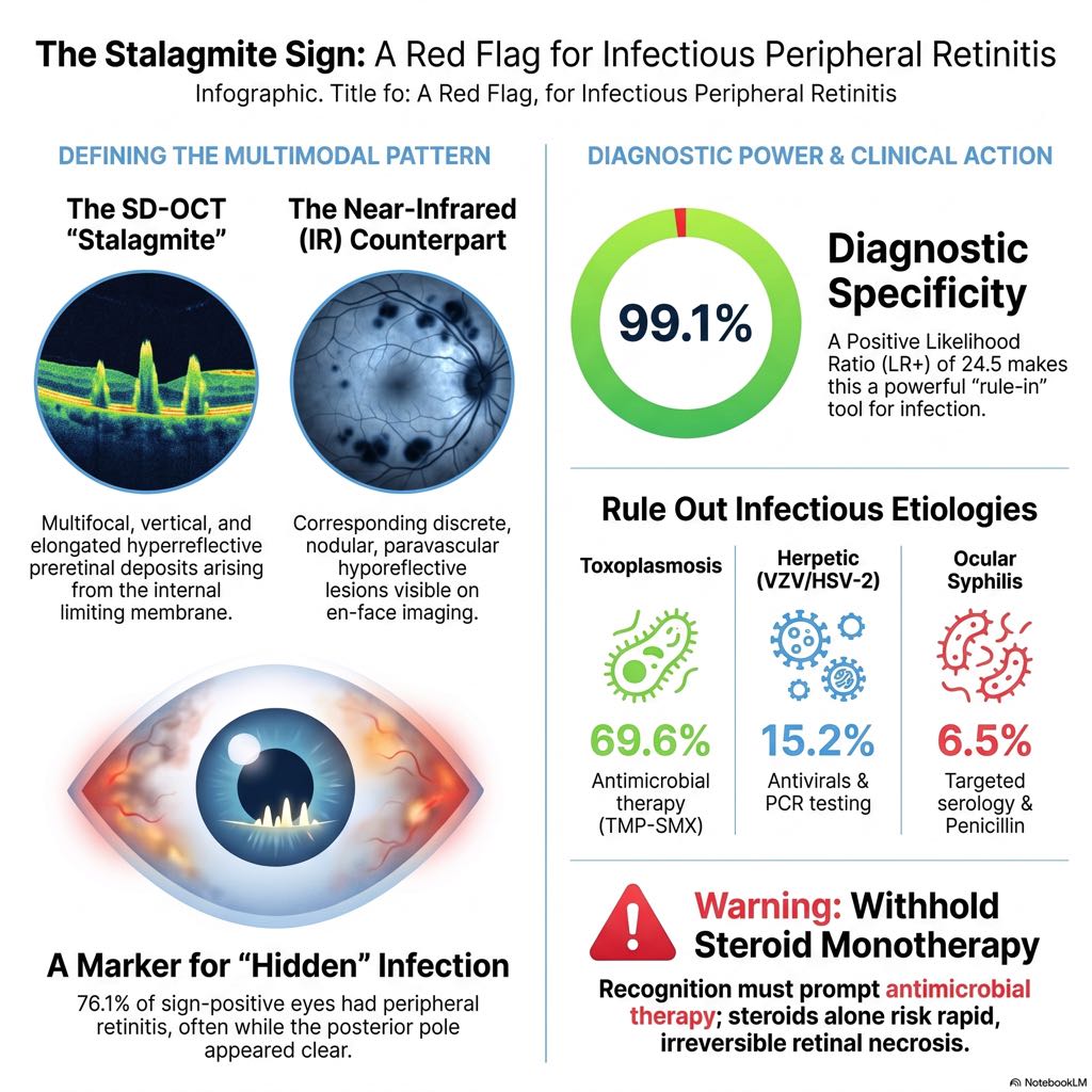
The Stalagmite Sign is not a solitary OCT finding; it is a unified multimodal pattern that demands precise spatial correspondence between two imaging channels. It is identified using confocal scanning laser ophthalmoscope (cSLO) platforms, such as the Heidelberg Spectralis, which provide simultaneous B-scans and near-infrared (IR) reflectance.
| SD-OCT B-scan Findings | Co-registered Near-Infrared (IR) Reflectance |
| Multifocal, elongated, and vertical hyperreflective preretinal deposits. These projections arise from the internal limiting membrane (ILM) and extend into the vitreous cavity. | Discrete nodular paravascular hyporeflective lesions. These appear as small, dark “nodules” along the retinal vessels on the en-face image. |
The name “Stalagmite” is morphologically apt, referencing the geological resemblance of these deposits as they taper upward from the ILM toward the posterior hyaloid. While the mean maximum height is approximately 108 ± 49 µm, the absolute height has not been shown to correlate with the location of the retinitis or final visual outcomes. Rather, it is the presence of the sign and its one-to-one spatial correspondence—where every hyperreflective spike on the B-scan aligns with a hyporeflective nodule on the IR image—that carries diagnostic weight.
The use of IR reflectance is technically significant. Because the cSLO IR light (820 nm) penetrates vitreous haze and inflammatory debris more effectively than the shorter wavelengths used in traditional fundus photography, these paravascular nodules are often “readily identifiable at a glance” even when the media is moderately hazy.
The Statistical Power: A “Rule-In” Biomarker
Data from a multicenter retrospective analysis of 406 eyes establishes the Stalagmite Sign as an exceptionally robust diagnostic tool. In clinical practice, we categorize it as a “Rule-In” biomarker—meaning its absence does not exclude infection, but its presence is nearly pathognomonic.
| Metric | Value (95% Confidence Interval) |
| Specificity | 99.1% (96.7–99.7%) |
| Positive Predictive Value (PPV) | 95.7% (85.5–98.8%) |
| Positive Likelihood Ratio (LR+) | 24.5 |
| Sensitivity | 22.9% (17.5–29.4%) |
| Negative Predictive Value (NPV) | 58.9% (53.7–63.9%) |
Clinical Decisiveness and Bayes’ Theorem
A Positive Likelihood Ratio (LR+) of 24.5 provides what we consider “clinically decisive” evidence. To understand the impact on clinical decision-making, we must apply Bayes’ Theorem to the pre-test and post-test probabilities of infection:
- In a patient with a 10% pre-test probability of infection, the presence of the Stalagmite Sign shifts the post-test probability to 73%.
- With a 30% pre-test probability, the probability of infection jumps to 91.3%.
- In a case where the clinical suspicion is 50%, the presence of the sign elevates the probability to 96.1%.
This dramatic shift necessitates an immediate change in management, specifically the withholding of steroids in favor of an infectious workup.
Pathophysiological Mechanism: The Immune Aggregate
The sign appears to be a physical manifestation of a high infectious burden. In conditions like toxoplasmic retinochoroiditis, the parasite disseminates hematogenously, establishing necrotic foci in the periphery. This triggers an intense local inflammatory response mediated by activated CD4+ T cells, Th1 cytokines, macrophages, and neutrophils.
These immune cells, along with fibrin and cellular debris, form “fibrin-inflammatory aggregates” at the vitreoretinal interface. While the primary infection may be in the periphery, these aggregates migrate or form along the posterior pole’s vascular arcades, where they adhere to the ILM. The Stalagmite Sign is, essentially, a visible marker of the high-energy immunological battle occurring elsewhere in the eye.
Morphological Distinction: Comparing Biomarkers
In the landscape of OCT biomarkers, the Stalagmite Sign must be distinguished from other described preretinal deposits (PDs):
- Invernizzi’s Oval Deposits: These are typically “oval” or rounded and are found on the inner retinal surface in classic toxoplasmosis. The Stalagmite Sign is distinguished by its vertical, elongated, “upright” configuration.
- Nakao’s Granular Deposits: Associated with HTLV-1, these are granular and perivascular but lack the tapering, vertical height of the stalagmite.
- “Pitchfork” or “Rain-cloud” Signs: While these follow the metaphorical naming conventions common in modern OCT nomenclature, the Stalagmite Sign is unique in its multimodal requirement—the IR reflectance “nodule” is as critical for the diagnosis as the B-scan “spike.”
Etiological Breakdown: What Pathogens are Hiding?
In the study cohort of 46 sign-positive eyes, an infectious etiology was confirmed in 95.7% of cases. The breakdown reveals that while toxoplasmosis is the primary driver, other pathogens are frequently involved:
- Ocular Toxoplasmosis (69.6%): The most common cause. These cases often involve hematogenous spread to the periphery.
- Herpetic Retinitis (15.2%): Including Varicella-Zoster Virus (VZV) and Herpes Simplex Virus (HSV-2). All herpetic cases in the series presented with peripheral retinitis.
- Ocular Syphilis (6.5%): Presenting as superficial retinal precipitates; the sign should prompt immediate RPR/TPHA testing.
- Fungal Endophthalmitis (4.3%): Specifically Candida albicans, typically presenting with dense vitritis.
The Peripheral Dissociation: The clinical “blind spot” is confirmed by the data: 76.1% of eyes with the Stalagmite Sign had active peripheral retinitis, while only 17.4% had involvement in the posterior pole. The sign is the clinician’s warning that the “quiet” posterior pole is a deception.
Clinical Management: The “Red Flag” Workflow
When the Stalagmite Sign is detected, we recommend a strict, 4-step algorithm:
- Comprehensive Peripheral Exam: Perform mandatory 360-degree indirect ophthalmoscopy with scleral depression. The sign indicates that a focus of necrotizing retinitis is almost certainly present in the periphery.
- Laboratory/Diagnostic Workup: Perform immediate aqueous humor paracentesis for multiplex PCR. In the study, PCR was 100% positive in diagnostically uncertain cases where the sign was present. This suggests the sign may be a surrogate for a higher pathogen load, making it more detectable in the anterior chamber.
- Withhold Steroids: This is a non-negotiable safety imperative. Corticosteroid monotherapy in the presence of this sign is contraindicated until infection is controlled with antimicrobials.
- Antimicrobial Initiation: Promptly start pathogen-specific therapy (valacyclovir, pyrimethamine/sulfadiazine, or antifungals).
Critical Mimics and Safety Differentiators
Clinicians must be wary of two specific mimics:
- Post-Silicone Oil Deposits: Deposits can appear after silicone oil removal following endophthalmitis surgery. The critical differentiator is the absence of vitritis. In the study, all infectious stalagmites were accompanied by vitritis, whereas silicone oil mimics were not and resolved spontaneously.
- Primary Vitreoretinal Lymphoma: In patients over 50 with chronic vitritis and negative PCR results, lymphoma must be considered. Diagnostic vitrectomy with IL-10/IL-6 ratios and MYD88 mutational analysis is warranted in these atypical, chronic cases.
Treatment Response and Visual Outcomes
The kinetics of the Stalagmite Sign’s disappearance serve as a visual surrogate for therapeutic success.
- Resolution: 69% of deposits resolve completely at 1 month, and 100% resolve by 3 months with proper antimicrobial therapy.
- Visual Acuity: Mean BCVA improved significantly from 0.64 ± 0.40 logMAR (~20/80) to 0.43 ± 0.45 logMAR (~20/50-20/60) over the follow-up period. The Friedman test p-value (<0.001) confirms the statistical significance of this improvement.
Surgical Intervention: Nearly 41.3% of eyes required surgical intervention, but the indications differed by etiology.
- Toxoplasmosis: Pars Plana Vitrectomy (PPV) was primarily indicated for vitreous condensation and associated epiretinal membranes (ERM). ERM peeling was performed in 90.9% of these cases, with 72.7% of patients gaining ≥2 lines of vision.
- Herpetic Retinitis: PPV was often necessitated by rhegmatogenous retinal detachment (RD), a common and severe sequela of herpetic necrosis.
Conclusion: Why the Stalagmite Sign Matters for Your Practice
Integrating the Stalagmite Sign into routine imaging review transforms the OCT from a macular screening tool into a powerful infectious disease sentinel. Its high specificity and decisive likelihood ratio provide the evidence needed to resist the “steroid trap” and pursue a life-saving—and sight-saving—infectious workup.
Key Takeaways for the Clinician:
- A Marker of Infectious Burden: The Stalagmite Sign is a high-specificity “red flag” for active infection, likely reflecting a significant systemic or intraocular pathogen load.
- The Peripheral Mandate: Its presence is an absolute indication for a meticulous peripheral retinal exam with scleral depression.
- The Safety Priority: When the sign is present, manage the patient as “infection first.” PCR testing is highly productive in these cases, and antimicrobial coverage must precede any consideration of corticosteroid therapy.
In modern retina practice, the simultaneous review of co-registered IR-reflectance and SD-OCT B-scans for these vertical hyperreflective spikes is no longer optional—it is a fundamental safety maneuver in the management of uveitis.

Reference:
Lasave, Andres F., et al. “The Stalagmite Sign: A Novel OCT-IR Biomarker for Peripheral Infectious Retinitis.” Am. J. Ophthalmol., vol. S0002-9394, no. 26, 21 July 2026, pp. 00404-06, doi:10.1016/j.ajo.2026.07.028.


