
1. Introduction: Beyond the Basics of Phototransduction
In the specialized architecture of the vertebrate retina, Arrestin-1—historically designated as S-antigen or the 48 kDa protein—functions as the definitive regulator of light-evoked signaling. While introductory models often characterize it as a binary “off-switch,” contemporary molecular retinology reveals a sophisticated molecular rheostat. Its primary mission is the quenching of rhodopsin signaling with sub-second kinetics, a necessity for maintaining high temporal resolution and ensuring the reproducibility of single-photon responses.
As specialists, we must reconcile the static “snapshots” provided by X-ray crystallography with the dynamic, solution-based realities of the living cell. This document synthesizes biochemical data and clinical pathology to provide an advanced perspective on Arrestin-1, moving from its role in signal desensitization to its broader impact on retinal metabolism and survival.
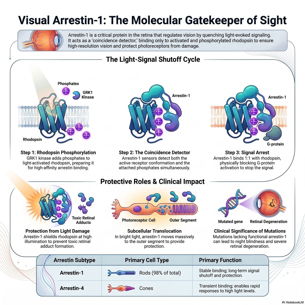
2. The Two-Step Dance of Desensitization
The cessation of rhodopsin signaling is governed by a “two-step mechanism of homologous desensitization,” involving the coordinated efforts of GRK1 (rhodopsin kinase) and Arrestin-1. This sequence ensures that rod photoreceptors operate as rheostats rather than simple on-off switches, allowing for a graded response to varying light intensities.
The Mechanism of Visual Arrest
| Step 1: Phosphorylation (GRK1) | Step 2: High-Affinity Binding (Arrestin-1) |
| Action: GRK1 multi-phosphorylates the C-terminus of light-activated rhodopsin (Rh^*). | Action: Arrestin-1 recognizes the phosphorylated, active receptor (P-Rh^*) and binds with high affinity. |
| Functional Outcome: Converts the active receptor into a high-affinity partner for arrestin, preparing it for signal shutoff. | Functional Outcome: Arrestin-1 physically shields the cytoplasmic side of the receptor, precluding further transducin activation. |
3. The “Coincidence Detector” and Structural Dynamics
Arrestin-1 acts as a “coincidence detector” through a sequential multi-site binding model. This ensures high-affinity engagement only when the protein encounters a receptor that is simultaneously active and phosphorylated. The molecular architecture involves a global conformational rearrangement triggered by specific sensors.
Molecular Trigger Points:
- The Polar Core (Arg175): This residue functions as a critical autoinhibitory gatekeeper. In the basal state, Arg175 is stabilized by an intramolecular interaction with Asp30, Asp296, and Asp303. Disruption of this core—either by rhodopsin-attached phosphates or specific mutations—relieves this inhibition.
- The Finger Loop (Residues 66–81): Functioning as the primary “activation sensor,” this loop transitions into an \alpha-helix upon engagement and inserts into the inter-helical cavity of active rhodopsin (Metarhodopsin\ II), stabilizing the active conformation.
- The C-Terminus: Basally, the C-terminus is anchored to the N-domain. High-affinity binding triggers its release, making it accessible to proteolysis and allowing the rest of the molecule to engage the receptor interface.
4. Clinical Correlations: When the Mechanism Fails
The molecular pathology of the retina is highly dependent on which component of the desensitization machinery is compromised. Mutations in RHO (rhodopsin), GRK1, and SAG (arrestin-1) produce distinct clinical phenotypes.
There is a notable “counterintuitive” discrepancy in clinical severity. In murine models, the knockout of GRK1 causes more significant abnormalities in single-photon responses than the SAG knockout. However, in humans, GRK1 deficiency typically manifests as Oguchi disease (stationary night blindness), whereas SAG deficiency frequently progresses to severe Retinitis Pigmentosa (RP). This suggests that Arrestin-1 performs vital morphological and protective functions that GRK1 does not.
A prime example of misfolding pathology is the dominant Cys147Phe mutation. This mutation triggers the unfolded protein response (UPR) due to the accumulation of defective protein, leading to photoreceptor demise even when a wild-type allele is present.
5. The Protective and Metabolic Roles of Arrestin-1
Arrestin-1 is expressed at nearly 1:1 stoichiometry with rhodopsin (approximately 0.8:1 in mice), reaching concentrations of ~2 mM. This abundance supports critical non-signaling functions.
Secondary Biological Functions:
- Light Damage Mitigation: By shielding rhodopsin from transducin at high illumination levels, Arrestin-1 protects the rod outer segments from light-induced damage.
- Retinal Flux Regulation: Arrestin-1 slows the release of all-trans-retinal from photobleached rhodopsin, preventing the formation of toxic retinal adducts. Fascinatingly, species like owls and deep-diving whales have acquired a Gln69Arg substitution in the finger loop through convergent evolution, an adaptation that enhances this ability to slow retinal flux.
- Metabolic Interaction: Arrestin-1 binds to and inhibits Enolase-1. Research involving the Glu362Gly/Asp363Gly mutant (which fails to bind Enolase-1) shows that regulating this glycolytic enzyme impacts lactate secretion and can slow retinal degeneration in Pro23His rhodopsin models.
6. Light-Induced Translocation: The Diffusion Mystery
One of the most dramatic physiological events is the light-induced translocation of Arrestin-1 from the inner to the outer segments. While early “Active Transport” hypotheses were popular, the energetics are impossible. Moving the massive abundance of Arrestin-1 via active transport would require 0.2–0.4 pmol of ATP; the weight of this ATP alone would exceed the weight of the rods themselves by 3 to 6 fold.
The movement is driven by diffusion and binding. In the dark, Arrestin-1 is “parked” in the microtubule-rich inner segment through interactions with tubulin (K_D \sim 40 \mu M). Upon illumination, the high-affinity sites on phosphorylated rhodopsin in the outer segment “pull” the protein across the cilium.
7. Structural Heterogeneity: Crystals vs. Reality
While crystallography provides essential models, solution-based DEER and NMR studies have corrected several misconceptions. Specifically, the “Solution Tetramer” differs from the “Crystal Tetramer.” This was proven by double alanine substitutions of Phe85 and Phe197 (bovine) or Phe86 and Phe198 (mouse), which disrupted the solution-based oligomers but not the crystal-based ones.
Specialists must also appreciate the concept of “different flavors” of the arrestin-receptor complex. Evidence from arrestin-2 interactions with PTH1R revealed 136 proximity points, proving mathematically that the complex exists as an equilibrium of many conformational states.
Solution NMR provides the definitive affinity values for these states:
- Rh^*: relatively low affinity (K_D > 150 \mu M).
- Dark P-Rh: higher affinity (K_D \sim 80 \mu M).
- P-opsin: very tight affinity (K_D \sim 800 nM), likely to suppress signaling “noise” from opsin before regeneration.
8. The “Cytotoxic Monomer” and Oligomerization
The self-association of Arrestin-1 into dimers and tetramers (which are incompetent for rhodopsin binding) serves a vital metabolic purpose. Oligomerization acts as a reservoir, ensuring a supply of monomeric arrestin is available for rapid binding while sequestering the rest. This is biologically critical, as an overabundance of the free monomer is known to be cytotoxic, inducing light-independent photoreceptor death.
9. Conclusion: Lessons for the Clinic
Arrestin-1 is far more than a “signal stopper.” It is a multifunctional scaffold involved in SNARE complex recycling (via NSF), metabolic regulation, and the sequestration of reactive retinoids. Its multifaceted nature explains why its absence is more deleterious than the loss of GRK1. For the clinician, understanding these molecular nuances—from the ATP requirements of translocation to the specific residues of the solution tetramer—is essential for navigating the complexities of hereditary retinal degenerations.

Reference:
Gurevich, Vsevolod V. and Eugenia V. Gurevich. “Visual arrestin-1: how did we learn what we know today about this protein?” Prog. Retin. Eye Res., vol. 113:101491., July 2026, doi:10.1016/j.preteyeres.2026.101491.


