
Rhegmatogenous retinal detachment (RRD) is a vision-threatening emergency that requires prompt and precise surgical intervention. For decades, the foundational principle of treating RRD has remained the same: find the retinal breaks and seal them. Whether a surgeon opts for a scleral buckle (SB), pars plana vitrectomy (PPV), or pneumatic retinopexy (PnP), the accurate preoperative localization of every single retinal break is the cornerstone of surgical success. Missing a tear, especially when placing a scleral buckle that must perfectly indent the break, often leads to surgical failure and recurrent detachment.
In recent years, Ultra-Widefield Imaging (UWFI), such as the Optos system, has revolutionized the way eye care professionals document and evaluate the retina. It is fast, patient-friendly, and provides a stunning panoramic view of the fundus. But a critical question remains for retina specialists: Can a single UWFI scan reliably replace the traditional, albeit uncomfortable, indirect ophthalmoscopy with scleral indentation?
According to a comprehensive 2026 study by Chou et al., published in Eye, the answer is a resounding no. Let’s dive into the data to understand the limitations of UWFI in detecting retinal breaks and explore how these findings should shape our clinical practice and surgical planning.
The Study: Putting UWFI to the Test
To evaluate the true sensitivity of UWFI, researchers at China Medical University Hospital conducted a retrospective analysis of 354 adult eyes with primary RRD that underwent pars plana vitrectomy (PPV).
To establish an airtight reference standard, the researchers didn’t just compare UWFI to clinical exams; they compared preoperative UWFI against intraoperative scleral indentation performed by experienced retina surgeons after complete vitrectomy and before fluid-air exchange. This intraoperative 360-degree inspection provides the most definitive confirmation of the presence, number, and location of all retinal breaks. The preoperative imaging protocol utilized a single, centrally steered Optos UWFI image captured after pharmacologic pupil dilation.
Breaks were categorized into two main groups:
- The Causative Break: The break judged by the surgeon to be most responsible for the detachment, factoring in size, traction, and subretinal fluid configuration.
- Additional Breaks: Any other breaks present in the detached or attached retina.
The Hard Numbers: How Much Are We Missing?
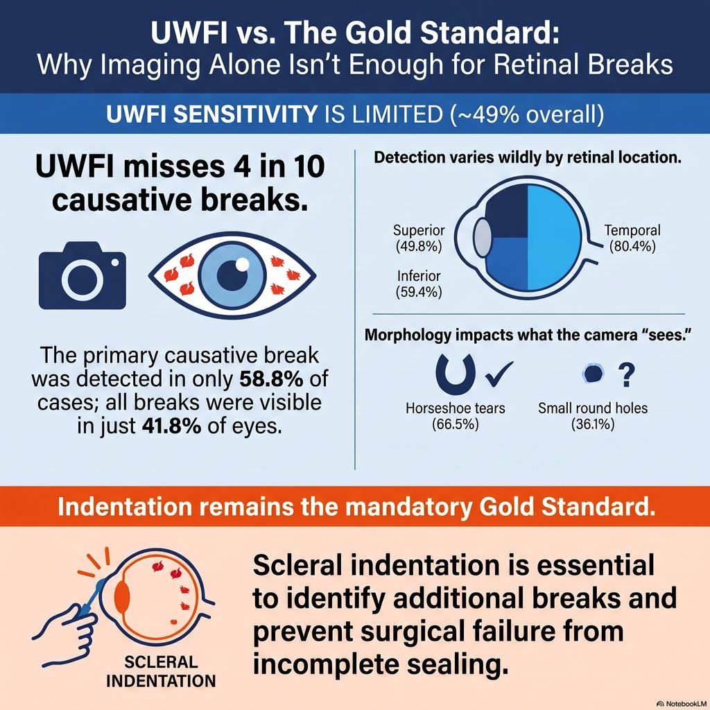
The results of the study serve as a humbling reminder of the limitations of our current imaging technology. When relying on a single, centrally steered UWFI image, the overall sensitivity for detecting all retinal breaks was only 49.1%. This means that more than half of all retinal breaks present in RRD are entirely invisible on standard UWFI.
When breaking down the data further, the detection rates were as follows:
- Causative Break Detection: The primary break responsible for the detachment was identified on UWFI in 208 of the 354 eyes (58.8%).
- Total Break Detection: All breaks (causative plus any additional breaks) were successfully identified on UWFI in only 41.8% of eyes.
Clinically, this is a massive pitfall. In eyes where the causative break was successfully detected on UWFI, nearly one-third of these patients had coexisting additional breaks that were completely missed by the imaging system. Relying solely on the UWFI would leave these secondary breaks untreated, almost guaranteeing postoperative complications and surgical failure.
Which Breaks Are Easier to See?
Not all retinal breaks are created equal on imaging. The study revealed distinct factors that make a break more or less likely to be captured on a UWFI scan.
1. Break Morphology: The shape and size of the tear heavily dictate its visibility.
- Horseshoe Tears: These were successfully detected 66.5% of the time. Their larger size and the presence of a tractional flap make them stand out against the detached retina.
- Round Holes: These were notoriously difficult to spot, with a detection rate of only 36.1%. Multivariable logistic regression showed that round holes were significantly less likely to be detected compared to horseshoe tears (adjusted odds ratio = 0.07).
- Giant Retinal Tears: Due to their massive size, 100% of giant tears in the study cohort were easily identified.
2. Patient Demographics and Anatomy: Patients whose causative breaks were detected on UWFI tended to be significantly younger (57.8 vs. 61.3 years) and were more likely to be phakic. Younger, phakic eyes generally possess clearer lenses and allow for better pupillary dilation. In contrast, older pseudophakic patients may suffer from posterior capsule opacification or anterior capsular phimosis, both of which severely degrade peripheral image quality. Furthermore, younger patients with RRD often have higher degrees of myopia and a more posterior vitreous base insertion, pulling tears into a more posterior (and thus more visible) location.
3. Extent of Detachment: Counterintuitively, a smaller detachment extent makes breaks easier to find on UWFI. Eyes with a 1- or 2-quadrant detachment had detection rates around 61-64%, while eyes with massive 3- to 4-quadrant detachments saw detection plummet to 40.3%. This is because extensive, highly bullous detachments frequently fold over themselves, obscuring underlying breaks, whereas smaller detachments present a flatter, more easily imaged topography.
The Geography of Detection: Quadrants and Clock Hours
Perhaps the most fascinating finding from this study is how drastically UWFI sensitivity varies depending on the anatomical location of the break.
- The Temporal Advantage: The temporal quadrant was by far the most reliably imaged area, boasting a 69.2% sensitivity for all breaks and an 80.4% sensitivity for causative breaks. Clock-hour analysis showed peak detection clustered precisely at 9 and 10 o’clock. Why? Anatomically, the temporal retina is less obstructed by facial features like the nose and brow. Furthermore, the ellipsoid mirrors and patient positioning algorithms of UWFI systems inherently favor the temporal periphery.
- The Inferior Blind Spot: The inferior quadrant is the Achilles’ heel of UWFI. It exhibited the lowest sensitivity, detecting only 33.0% of total breaks and a dismal 22.9% of additional breaks. Clock hours 5 and 6 o’clock represented the absolute lowest points of detection. Eyelashes, drooping eyelids, and the Bell’s phenomenon (upward rolling of the eyes during imaging) frequently shadow the inferior retina.
- Superior and Nasal Quadrants: These areas fell in the middle, with sensitivities of 45.2% and 45.9% for total breaks, respectively. Bullous superior detachments can easily be obscured by upper lid lashes.
Technical Refinements vs. Clinical Reality
It is worth noting that this study evaluated standard, single, centrally steered UWFI. Some proponents of widefield imaging argue that utilizing peripheral eye steering (capturing images while the patient looks in 4 to 8 different directions) or using automated image montages can dramatically increase peripheral capture.
Studies utilizing the Zeiss Clarus system or multi-image Optos montages have indeed reported higher sensitivities (upward of 85%). However, executing a flawless multi-image peripheral montage requires a highly cooperative patient and significantly more technician time. In a busy real-world retina clinic, capturing a single centrally steered image is the standard pragmatic approach. Therefore, this study accurately reflects the baseline sensitivity you can expect from routine, high-volume clinical UWFI use.
📚 BOARD EXAM-SPECIFIC HIGHLIGHTS
For ophthalmology residents and optometry students preparing for board exams, the interplay between retinal detachments and imaging modalities is high-yield material. Memorize these key pearls derived from this and similar studies:
- The Gold Standard Never Changes: Preoperative indirect ophthalmoscopy with scleral indentation remains the absolute gold standard for detecting retinal breaks in RRD. UWFI is an adjunct, not a replacement.
- Beware the Inferior Quadrant: On board questions regarding UWFI (like Optos) limitations, remember that the inferior quadrant (5 and 6 o’clock) has the lowest sensitivity for break detection due to interference from eyelids and eyelashes.
- The Temporal Advantage: The temporal retina (9 and 10 o’clock) is the easiest to image on UWFI because the optical pathway is not obstructed by the nose or brow.
- Morphology Matters: Horseshoe tears are vastly more likely to be detected on UWFI than atrophic round holes. If a vignette describes a missed break on UWFI, it is highly likely to be a small round hole or located inferiorly.
- Surgical Implications for Scleral Buckles (SB): Missing a break is catastrophic for Scleral Buckling. The buckle must support the break. Because UWFI misses roughly 60-80% of additional breaks, relying on imaging alone for SB planning is contraindicated.
- Phakic vs. Pseudophakic: Phakic eyes generally yield better UWFI quality in RRD patients than pseudophakic eyes, often due to posterior capsular opacities obscuring the periphery in the latter.
Conclusion
Ultra-widefield imaging is an undeniably powerful tool. It provides incredible preoperative documentation, assists in patient education, and gives surgeons a rapid, high-resolution map of the primary detachment architecture. It is particularly adept at confirming temporal causative breaks and large horseshoe tears.
However, technology has not yet surpassed the human touch. With a total break detection rate hovering just below 50%, a normal or “single-break” UWFI scan is a dangerous false comfort. Relying exclusively on this imaging modality invites surgical failure through missed secondary breaks, particularly in the obscured inferior retina.
For the modern retina specialist, the workflow is clear: use the UWFI to document the macro-architecture of the detachment, but always pick up your 20-diopter lens and scleral depressor to find the truth. UWFI complements, but will never replace, a thorough, dynamic clinical examination.

Reference: Chou, H.-C., Su, Y.-H., Lu, J.-Y., Altamirano, F., Patel, N. A., Huang, Y.-T., & Chen, S.-N. (2026). Sensitivity of ultra-widefield fundus photography for retinal break localisation in primary rhegmatogenous retinal detachment. Eye. https://doi.org/10.1038/s41433-026-04608-9


