
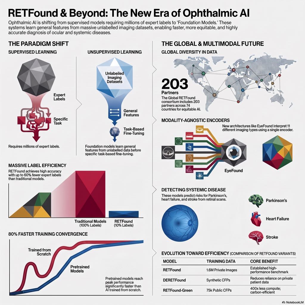
Artificial intelligence (AI) has dominated medical headlines for years, but ophthalmology is currently experiencing a profound paradigm shift. We are moving away from traditional, narrow deep learning algorithms designed for single tasks toward incredibly versatile, generalizable systems known as foundation models. For retina specialists, these models promise to revolutionize diagnostic workflows, transform community screening, and unlock new insights into systemic health through the eye.
What is a Foundation Model? Coined in 2021, the term “foundation model” describes a massive AI architecture trained on vast quantities of diverse, unlabelled data that can be adapted to a myriad of downstream clinical tasks.
Traditional deep learning relies heavily on supervised learning, which requires thousands of meticulously expert-labeled images (like grading diabetic retinopathy severity) to teach the AI a specific task. This process is immensely expensive, time-consuming, and limits the AI to that one specific function.
Foundation models bypass this bottleneck using Self-Supervised Learning (SSL). Because unlabelled retinal images accumulate by the millions in routine clinical care, SSL taps into this unused data. The model trains itself by playing a complex game of “fill-in-the-blank.” For example, using a generative approach like masked autoencoding, the system hides 75% to 80% of an optical coherence tomography (OCT) scan or color fundus photograph (CFP) and forces the AI to reconstruct the missing anatomical structures. Alternatively, contrastive learning teaches the model to mathematically group augmented views of the same image while pushing apart unrelated images.
Underpinning these models is the Transformer architecture. Unlike traditional convolutional neural networks (CNNs) that focus on local pixel neighborhoods, transformers use “attention mechanisms” to understand the global context of an image—meaning the AI can map the relationships between a macular lesion and peripheral vascular changes simultaneously.
The resulting foundation model possesses incredible label efficiency, the ability to generalize across different populations, and a drastically reduced computational burden when fine-tuned for a specific task.
The Evolution of Ophthalmic Foundation Models The journey of foundation models in ophthalmology has occurred in four distinct phases:
1. Natural-Image Models Early efforts attempted to apply models trained on millions of general internet images (like dogs, cars, and landscapes) to medical tasks. Systems like the Segment Anything Model (SAM) and REMEDIS showed preliminary promise. However, generalist models often struggled to adapt to the fine-grained, specialized anatomical textures of the retina, such as branched retinal vessels, without heavy domain-specific fine-tuning.
2. Unimodal Retinal Models The breakthrough came with the development of domain-specific models like RETFound, published in Nature in 2023. RETFound was pre-trained on 1.6 million unlabelled CFPs and OCTs from Moorfields Eye Hospital. It learned the specific visual language of the retina.
When adapted to specific tasks, RETFound consistently matched or outperformed traditional supervised AI in diagnosing intrinsic ocular diseases like diabetic retinopathy, glaucoma, and macular degeneration. Furthermore, it unlocked the field of oculomics—predicting systemic health risks from retinal imaging. RETFound successfully predicted the 3-year risk of heart failure, myocardial infarction, ischemic stroke, and Parkinson’s disease directly from retinal scans. Crucially, it demonstrated unmatched label efficiency, achieving expert-level performance in heart failure prediction using only 10% of the normally required labeled data, and reducing training time by 80%.
Newer variants have further optimized this framework. DERETFound utilizes generative AI to create synthetic CFPs for training, matching RETFound’s performance using 50% less human-annotated data during fine-tuning. Meanwhile, RETFound-Green applies a novel “token reconstruction” method, building a highly capable model on consumer-grade hardware for a fraction of the computational and environmental cost.
3. Multimodal Foundation Models In the real world, retina specialists rarely rely on a single imaging modality. The next frontier in foundation models was integrating complementary data streams. Early multimodal models like VisionFM trained separate encoders for up to 8 different modalities (CFP, OCT, ultrasound, MRI, etc.).
More advanced iterations like URFound and EyeFound use a single, shared “modality-agnostic” encoder. By training the exact same neural network on CFPs, OCTs, fluorescein angiography (FFA), and indocyanine green angiography (ICGA), the model learns universal cross-modal features. EyeFound, trained on 2.78 million images across 11 modalities, significantly improved diagnostic accuracy on complex multimodal datasets compared to unimodal models.
4. Vision-Language Models (VLMs) The most cutting-edge models align retinal images with clinical text. Models like FLAIR, RetiZero, and EyeCLIP use contrastive learning to map a patient’s fundus image and their corresponding unstructured clinical diagnostic report into a shared AI space.
This unlocks a superpower for the clinician: Zero-shot inference. Because the model understands the semantic relationship between textual concepts and visual features, you can ask the AI to identify a rare condition (e.g., “retinitis pigmentosa”) without ever having explicitly trained the model on labeled examples of that disease. RET-CLIP, trained on nearly 200,000 patients with left/right eye CFPs matched to free-text clinical reports, achieved state-of-the-art multi-disease classification by learning the holistic clinical reasoning embedded in human-authored notes.
The Global RETFound Initiative: Solving the Bias Problem Despite these massive advancements, a dangerous flaw plagues medical AI: models are usually trained on geographically and demographically narrow data. A model trained strictly on urban European populations may underperform or perpetuate inequities when deployed in South America or Africa.
To solve this, a consortium of over 100 study groups across 65 countries has launched the Global RETFound initiative. This collaboration is compiling an unprecedented dataset of over 100 million color fundus photographs covering every continent except Antarctica.
To navigate strict international data privacy laws and infrastructure barriers, the consortium utilizes a highly innovative two-pronged data-sharing framework:
- Synthetic Data Generation: Institutions with technical capabilities receive a Docker container to locally fine-tune a generative AI model on their own anonymized patient data. The hospital then shares only the mathematical model weights (not the patient images) with the central hub. The central hub uses these weights to generate millions of synthetic, demographically representative images that preserve crucial anatomical features without compromising privacy.
- Secure Real Data Sharing: For partners lacking local GPU hardware, de-identified CFPs and metadata are securely uploaded directly to a central cloud.
The resulting model, Global RETFound Beta, is already outperforming existing tools in detecting ocular diseases, predicting age, and extracting biomarkers. Best of all, it will be released as an open-access resource, establishing a new international benchmark for fairness and equity in medical AI.
Clinical Implications for the Retina Specialist Foundation models are not designed to replace specialists; they are designed to augment clinical capacity. Here is what this means for the future of your practice:
- Workflow Optimization: Models can act as autonomous triage agents, identifying high-risk scans (like aggressive macular fluid or imminent neovascularization) and bumping them to the top of your review queue. Dual-headed models that output an “uncertainty score” can automatically flag ambiguous, non-diagnostic scans for human verification.
- Rare Disease Identification: Vision-Language models can serve as a real-time diagnostic partner. By utilizing zero-shot learning, the AI can cross-reference an atypical fundus presentation against hundreds of rare disease concepts derived from global literature, helping you prioritize differential diagnoses.
- Opportunistic Systemic Screening: Routine retinal imaging will increasingly be used to flag patients at high risk for undiagnosed cardiovascular disease, diabetic vascular complications, or dementia, allowing you to refer them to primary care or neurology long before systemic symptoms manifest.
- Democratized Research: If you have a small dataset of 500 images for a niche pathology, you no longer need massive computing power to build an AI. You can simply download an open-source model like RETFound and “fine-tune” it on your small dataset over a few hours to achieve expert-level diagnostic tools.
Challenges and the Road Ahead While the potential is vast, integrating these models into real-world healthcare requires caution. We must remain vigilant regarding bias; while Global RETFound is a massive step forward, AI can still reinforce harmful correlations. Data privacy is a persistent concern, as highly complex foundation models have occasionally been shown to “memorize” and regurgitate fragments of their training data. Finally, the “black box” nature of transformers creates regulatory and explainability hurdles; for a model to be trusted, it must be able to reference evidence-based sources and explain why it made a specific clinical recommendation.
Conclusion We are witnessing a monumental leap in computational ophthalmology. By leveraging self-supervised learning, transformers, and globally inclusive datasets, foundation models are transitioning AI from single-task algorithms to robust, multi-modal diagnostic partners. For retina specialists, this era promises more efficient workflows, deeper clinical insights, and fundamentally more equitable patient care.

Reference List:
- Chia MA, Antaki F, Zhou Y, Turner AW, Lee AY, Keane PA. Foundation models in ophthalmology. Br J Ophthalmol. 2024;108:1341–1348.
- Tham YC, Goh JHL, Nderitu P, Zhou Y, Ran AR, et al. Building the world’s first truly global medical foundation model. Nature Medicine. 2025.
- Nderitu P, Harp MD. ARVO 2026: Global RETFound study. Ophthalmology Times. May 14, 2026.
- Moorfields Eye Hospital. Quest to build the first medical AI foundation model with globally representative data. (Press Release). September 8, 2025.
- UCL Global. Using retinal images to predict disease across the world. November 8, 2025.
- Gharbi K, van Wijngaarden P, Hadoux X. Foundation models for ophthalmic imaging. Survey of Ophthalmology. 2026;71:1129–1147.


