
Central serous retinopathy (CSR) continues to be a complex condition in retinal practice. First extensively described by Gass as a serous detachment of the neuroepithelium driven by underlying choroidal pathology and sub-RPE leakage, CSR presents as macular or extramacular subretinal detachments frequently accompanied by pigment epithelial detachments (PEDs).
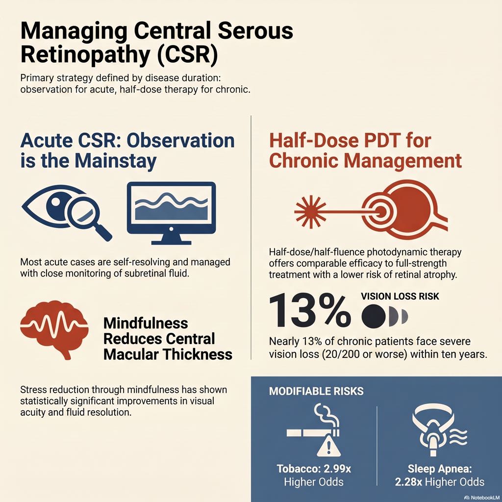
While acute CSR often resolves spontaneously with minimal visual consequence, chronic CSR presents a significant management challenge. For roughly 13% of patients with chronic CSR, best-corrected visual acuity (BCVA) deteriorates to 20/200 or worse within a 10-year follow-up period. Affecting nearly 6 in 100,000 individuals—with a distinct male predilection of 5.7 to 10 times that of females—CSR requires nuanced, evidence-based management.
This comprehensive review details the most recent advancements in genomics, imaging, risk factor profiling, and therapeutic interventions for retina specialists.
Decoding the Pathophysiology: Genomics and the AMD Connection
Recent genome-wide association studies (GWAS) have profoundly expanded our understanding of CSR’s underlying molecular mechanisms, particularly its overlap with age-related macular degeneration (AMD).
- Complement Pathway Dynamics: Initial GWAS identified variants in the CFH gene expression linked to CSR risk. Interestingly, while the complement pathway is often upregulated in AMD, targeted studies note an inverse relationship, with pathway downregulation observed in CSR.
- Shared Genetic Loci: Despite divergent complement activity, CSR and wet AMD share multiple genetic loci, including WBP1L, GATA5, CFH, C2/FB, TNFRSF10A, NOTCH4, and ARMS2.
- The Vascular Hyperpermeability Hypothesis: The PTPRB gene has emerged as a key player. Loss of PTPRB disrupts the tyrosine kinase receptor Tie-2 and VE-cadherin expression, both essential for endothelial cell integrity and angiogenesis. Underscoring a systemic vascular etiology, the PTPRB variant increasing CSR risk is also linked to the development of varicose veins.
- Hormonal Links: Another critical finding is the CDH7 gene, whose expression is associated with cortisol levels. This variant induces a higher odds ratio of CSR in male patients, providing a robust molecular basis for two of the most well-known clinical risk factors: male sex and exogenous steroid use.
Diagnostic Innovations in Multimodal Imaging
While traditional optical coherence tomography (OCT) remains the workhorse for diagnosing subretinal and sub-RPE fluid, advancements in multimodal imaging are refining our approach to complications like macular neovascularization (MNV/CNV).
- Optical Coherence Tomography Angiography (OCTA): OCTA has largely supplanted dye angiography as a primary tool for detecting neovascularization in CSR. It boasts excellent sensitivity (92.9%) and specificity (99.4%) while being non-invasive.
- Enhanced Depth Imaging (EDI-OCT): EDI-OCT consistently demonstrates persistent choroidal thickening in both acute and chronic CSR.
- Artificial Intelligence: Deep learning models are being developed to diagnose CSR using OCT findings, achieving near 100% accuracy, sensitivity, and specificity in early studies. However, widespread clinical adoption awaits validation on larger, more diverse training datasets.
Re-evaluating Clinical Risk Factors
While corticosteroids and “Type A” personalities are classic associations, recent large-scale meta-analyses have solidified new systemic risk factors:
- Obstructive Sleep Apnea (OSA): A massive meta-analysis of over 332,000 patients revealed that individuals with CSR have 2.28 times higher odds of having a concurrent OSA diagnosis. Retina specialists should consider OSA screening, given its systemic health implications and impact on the choroid.
- Tobacco Use: Smoking is a heavily modifiable risk factor. Data from nearly 28,000 CSR patients demonstrates that tobacco users have a 2.99-fold increased odds of developing the disease. This is likely due to the dose-dependent reduction in the choroidal vascularity index observed on OCT in smokers.
- Predicting Chronicity: When triaging patients, age is a significant intrinsic risk factor. A logistic regression model developed by the Macula Society CSCR Study Group found a 22.3% increased odds of developing chronic CSR for every advancing year of age. Patients presenting with poorer initial visual acuity, prolonged symptoms, reduced neurosensory detachment height, and lower central macular thickness (indicative of defective RPE) are also at higher risk of chronicity.
Evidence-Based Management of CSR in 2026
Observation remains the cornerstone of management for acute CSR, alongside the mitigation of known risk factors. However, for recurrent or chronic cases, intervention is often required. Recent trials have drastically shifted the therapeutic landscape.
What Does Not Work: NSAIDs and Long-term MRAs
- Topical NSAIDs: Despite their utility in inflammatory cystoid macular edema, a meta-analysis of 1,001 eyes showed no statistically significant benefit in visual acuity or fluid resolution at 3 months with topical NSAIDs.
- Mineralocorticoid Receptor Antagonists (MRAs): While early RCTs suggested spironolactone and eplerenone could rapidly reduce subretinal fluid (SRF) at 1 month, a large meta-analysis (646 eyes) debunked their long-term efficacy. There is no significant difference in BCVA at up to 1-year follow-up compared to observation. In fact, patients in the observation cohort had a higher rate of SRF resolution at one year. Given the risks of hyperkalemia and gynecomastia, the routine use of MRAs for chronic CSR is falling out of favor.
A Novel Approach for Acute CSR: Mindfulness
Given the strong association between CSR, psychological stress, and cortisol elevation, a randomized control trial evaluated mindfulness-based stress reduction. Patients practicing mindfulness had significantly better BCVA and reduced central macular thickness at 1, 3, and 6 months compared to controls, entirely avoiding pharmacological risks.
The Gold Standard: Photodynamic Therapy (PDT)
For chronic CSR, PDT remains a highly effective intervention. Activation of verteporfin induces choroidal vascular occlusion, reducing the choroidal hyperpermeability characteristic of the disease.
- Dosing Protocol: Full-dose/full-fluence PDT carries a dangerous risk of juxtafoveal RPE atrophy and permanent visual decline. Recent meta-analyses confirm that half-dose or half-fluence PDT achieves equivalent BCVA improvement and SRF resolution to full-strength treatment, but with a vastly superior safety profile. Low-dose PDT was ranked highest for 1-year BCVA outcomes in a recent large network meta-analysis.
Alternatives to PDT: Laser Therapies
In an era of recurrent verteporfin shortages, laser therapy serves as a viable alternative, though direct comparisons in the PLACE trial favored PDT for complete SRF resolution and BCVA improvement.
- Subthreshold/Micropulse Laser: Nanosecond subthreshold and microsecond pulsing lasers can effectively reduce SRF (up to 85% resolution at 1 year in some trials), though statistically significant BCVA improvements are less consistent.
- Navigated Focal Laser: For CSR complicated by small, extrafoveal CNV, navigated focal thermal laser is highly precise. In a study of 16 eyes (many resistant to anti-VEGF and PDT), complete fluid resolution was achieved in 100% of eyes at 1.1 months, with no recurrences at 1 year.
The Future of Pharmacotherapy: Targeted Anti-VEGF
Standard anti-VEGF agents generally do not significantly improve BCVA in uncomplicated chronic CSR, though they may reduce macular thickness and are essential if CNV is present. However, targeted mechanisms offer new hope:
- Faricimab (Targeting Tie-2): Motivated by GWAS data showing Tie-2 loss in CSR, faricimab (which inhibits Ang-2 to increase Tie-2 expression) was evaluated in a retrospective series. Fourteen out of 16 eyes with chronic, treatment-resistant CSR showed improved central macular thickness, indicating strong biological plausibility.
- High-Dose Aflibercept: In at least one documented case of intractable CSR failing PDT, oral eplerenone, and standard anti-VEGF regimens, a single off-label injection of high-dose intravitreal aflibercept prompted complete resolution of subretinal fluid.
Conclusion
The management of Central Serous Retinopathy requires a highly tailored approach. For acute presentations, non-invasive strategies like observation, risk factor modification (smoking cessation, OSA management), and stress-reduction techniques remain paramount. For chronic disease, half-dose/half-fluence PDT is the benchmark for safety and efficacy. As our genomic understanding deepens—particularly regarding the Tie-2 pathway—novel therapies like faricimab may soon offer targeted, molecular-level interventions for our most challenging chronic CSR cases.

Reference: Reeves, M.-G. R., & Talcott, K. E. (2026). Central serous retinopathy: update on disease understanding and treatment. Current Opinion in Ophthalmology, 37(3), 143–148. (Uploaded Source: “3-Central serous retinopathy update on disease understanding and treatment..pdf”).


