
As retina specialists, we have witnessed a remarkable evolution in the management of rhegmatogenous retinal detachment (RRD). Since Jules Gonin first identified the retinal break as the primary etiology in 1920, our surgical success rates have climbed from under 60% in the 1930s to approximately 90% today. Yet, despite our technical mastery of the “cure,” a critical question remains: Are we doing enough to prevent the disease in the first place?
The Problem with Focal Prophylaxis
The standard approach to prophylaxis has long been reactive, focusing on “high-risk” lesions like lattice degeneration identified during a peripheral exam. However, evidence suggests this strategy is fundamentally flawed. Studies show that in up to 90% of RRD cases, the causative break occurs in areas that appeared normal during previous examinations.
Furthermore, for a patient presenting with an RRD in one eye, the fellow eye carries a significant ~4% annual risk of developing its own detachment. Relying solely on the identification of focal pathology ignores the underlying pathology common to all RRDs: dynamic vitreoretinal (VR) traction.
Revisiting Laser Cerclage
While cryopexy is an established prophylactic tool, its associated morbidity and risk of intraocular inflammation—which can lead to proliferative vitreoretinopathy (PVR)—make it a less-than-ideal choice for widespread prevention.
Laser retinopexy, conversely, offers a safer alternative. The key to its success lies in moving beyond focal treatment toward 360-degree laser cerclage. Historically, laser cerclage earned a poor reputation due to inadequate application; however, when performed correctly, it is a highly effective “shield” for the peripheral retina.
Technique: The “Proper” Cerclage
To be effective, laser cerclage must address the entire “danger zone” where breaks typically form: the area between the ora serrata and the equator. A proper procedure involves:
- Comprehensive Coverage: Applying approximately 1,200 moderate burns in an emmetropic eye.
- Anatomic Landmarks: Sparing the 3 and 9 o’clock meridians to protect the long ciliary nerves.
- Full Extension: Ensuring treatment reaches the “ora secunda” (the posterior border of the treated retina) all the way to the ora serrata.
This can be performed as an outpatient procedure via indirect ophthalmoscope or during vitrectomy if scleral indentation allows for adequate peripheral reach.
Risk vs. Reward
The primary concern often cited is the risk of macular pucker formation, which occurs in approximately 1% of cases. While not zero, both the incidence and severity of a pucker are significantly lower than the devastating consequences of an actual RRD.
A New Standard of Counseling
Given that laser cerclage has been proven safe and effective, it is time to shift our counseling paradigm. Every patient presenting with an RRD should be informed of the ~4% annual risk to their fellow eye and offered the option of prophylactic laser cerclage. By providing patients with the data on both the risks of the procedure and the risks of observation, we empower them to choose true prevention over a future of potential surgery.

References
- Kuhn F, Aylward B (2014) Rhegmatogenous retinal detachment: a reappraisal of its pathophysiology and treatment. Rev Ophthalmic Res 51:15–31.
- Rezaei KA, Abrams GW (2005) The history of retinal detachment surgery. In: Kreissig I (ed) Primary retinal detachment. Springer, Berlin, Heidelberg, pp 1–25.
- Folk JC, Bennett SR, Klugman MR, Arrindell EL, Boldt HC (1990) Prophylactic treatment to the fellow eye of patients with phakic lattice retinal detachment: analysis of failures and risks of treatment. Retina 10:165–169.
- Wilkinson CP (2000) Evidence-based analysis of prophylactic treatment of asymptomatic retinal breaks and lattice degeneration. Ophthalmology 107:12–15.
- Morris RE, Kuhn F, Sipos T (2022) Preventing Retinal detachment: where are we? Implications from Stickler Syndrome. Clin Ophthalmol 16:4315–4321.
- Kuhn F, Morris R (2024) Rhegmatogenous retinal detachment: time to consider real prevention. Graefe’s Archive for Clinical and Experimental Ophthalmology. https://doi.org/10.1007/s00417-024-06725-1.
Risk Stratification in the Fellow Eye: Managing Lattice Degeneration After Primary RRD
For retina specialists, the discovery of lattice degeneration in the fellow eye of a patient who has just experienced a primary rhegmatogenous retinal detachment (RRD) presents a recurring clinical dilemma: to treat or to monitor? A recent large-scale retrospective study provides critical data to help refine our risk stratification and identify which patients benefit most from prophylactic intervention.
The Impact of Contralateral History
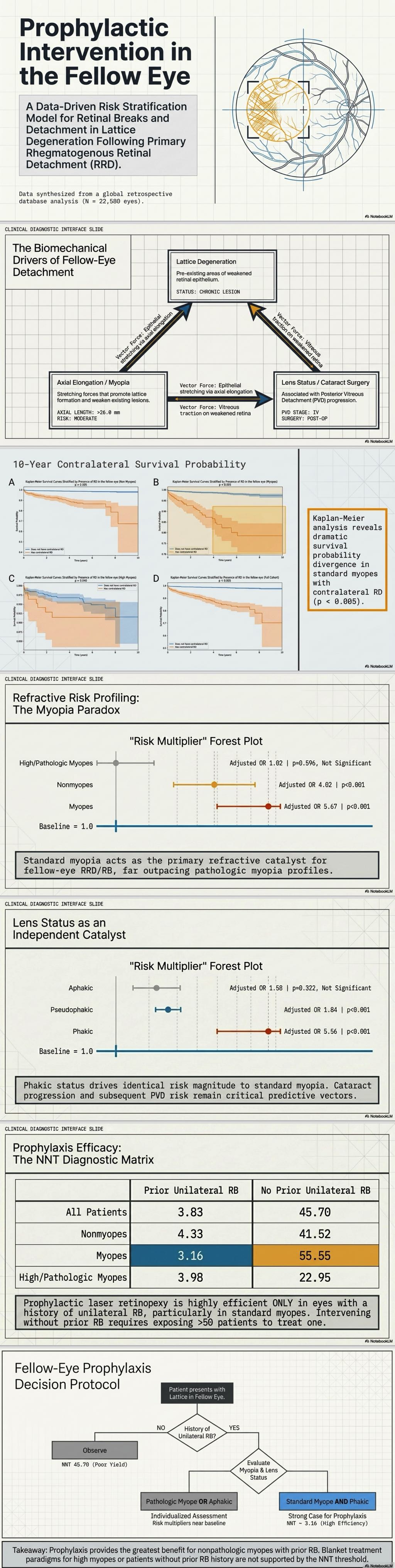
The most significant predictor of future pathology in a fellow eye with lattice is a history of RRD or retinal breaks (RB) in the first eye. The study found that patients with a history of contralateral RRD have an adjusted odds ratio (OR) of 4.47 for developing a similar event in the fellow eye.
Key Risk Factors for Progression
The risk is not uniform across all patients with lattice. Key variables include:
- Refractive Status: Non-pathologic myopes with lattice in the fellow eye are at particularly high risk (Adjusted OR 5.67). Interestingly, while axial elongation promotes lattice formation and weakens existing areas, the risk profile for high/pathologic myopes in this study was less pronounced (Adjusted OR 1.02), though still clinically significant.
- Lens Status: Phakic eyes with lattice were found to have a much higher risk of progression (Adjusted OR 5.56) compared to pseudophakic eyes (Adjusted OR 1.84). This may be influenced by the progression of posterior vitreous detachment (PVD) following cataract surgery, which remains a known risk factor for RRD.
- Demographics: The study noted higher incidences of these events in males (58.08%) and patients in the 50–69 age bracket.
Quantifying the Benefit: Number Needed to Treat (NNT)
The study utilizes NNT to evaluate the efficiency of prophylactic laser retinopexy or cryopexy. The data strongly suggests that prophylaxis is most effective when a prior event has already occurred in the first eye.
| Clinical Scenario | NNT to Prevent RRD | NNT to Prevent RB |
|---|---|---|
| Prior Contralateral Event | 4.63 | 3.83 |
| No Prior Contralateral Event | 43.29 | 45.70 |
Within the high-risk group (prior RRD), non-pathologic myopes showed the greatest benefit from treatment, with an NNT of only 3.59 to prevent an RRD.
Clinical Takeaways
Risk stratification is essential before proceeding with prophylactic retinopexy. Patients with non-pathologic myopia and a history of RRD in the first eye represent the highest-yield candidates for treatment. While these findings offer a robust framework for decision-making, prospective studies remain necessary to fully validate these interventions in various clinical settings.
Key findings regarding high myopes compared to non-myopes include:
- Higher Incidence of Events: In the study’s cohort, 11.05% of high/pathologic myopes experienced a fellow eye event (RRD or RB), compared to 6.31% of non-myopes.
- Greater Benefit from Prophylaxis (Lower NNT): For patients without a prior contralateral event, the Number Needed to Treat (NNT) to prevent one RRD was significantly lower for high myopes (14.21) than for non-myopes (39.54). A lower NNT indicates that the intervention is more “efficient” because the baseline risk in that group is higher.
- The “Risk Profile” Nuance: While high myopes are at a higher baseline risk, the study found that a prior history of RRD in the first eye was a much less significant predictor of a future event for them (Adjusted Odds Ratio of 1.02, which was not statistically significant) compared to non-myopes (Adjusted OR of 4.02).
In summary, while high myopes have a higher overall incidence of these events, the relative increase in risk triggered by a prior detachment in the other eye is less pronounced for them than it is for non-myopes or non-pathologic myopes.
References
- Ludwig, C., et al. The Risk of Retinal Breaks or Detachment in Fellow Eye with Lattice Degeneration after Primary Rhegmatogenous Retinal Detachment. Retina. 2026;46(7):1170-1175.
- Olsen TW, et al. Posterior vitreous detachment, retinal breaks, and lattice degeneration preferred practice pattern. Ophthalmology. 2020;127:P223–P258.
- Byer NE. Lattice degeneration of the retina. Surv Ophthalmol. 1979;23:213–248.
- Qureshi MH, Steel DH. Retinal detachment following cataract phacoemulsification—a review of the literature. Eye. 2020;34:616–631.


