Vision on the Brink: How Swept-Source OCTA Uncovered Choroidal Infarcts in HELLP Syndrome
🧠 Clinical Background
HELLP syndrome (Hemolysis, Elevated Liver enzymes, Low Platelets) is a severe complication of pregnancy, often associated with preeclampsia and eclampsia.
Ophthalmic manifestations include serous retinal detachments, retinal hemorrhages, cotton-wool spots, and choroidal ischemia.
👁️ Case Presentation
Patient: 36-year-old pregnant woman at 26 weeks gestation.
Symptoms: Bilateral blurry vision, abdominal pain, petechiae, and severe hypertension (BP 200/130 mmHg).
Initial Fundus Exam: Subretinal fluid and Elschnig spots in the right eye.
OCT Findings: Outer retinal disruption, hyperreflective deposits on Bruch’s membrane, thickened choroid.
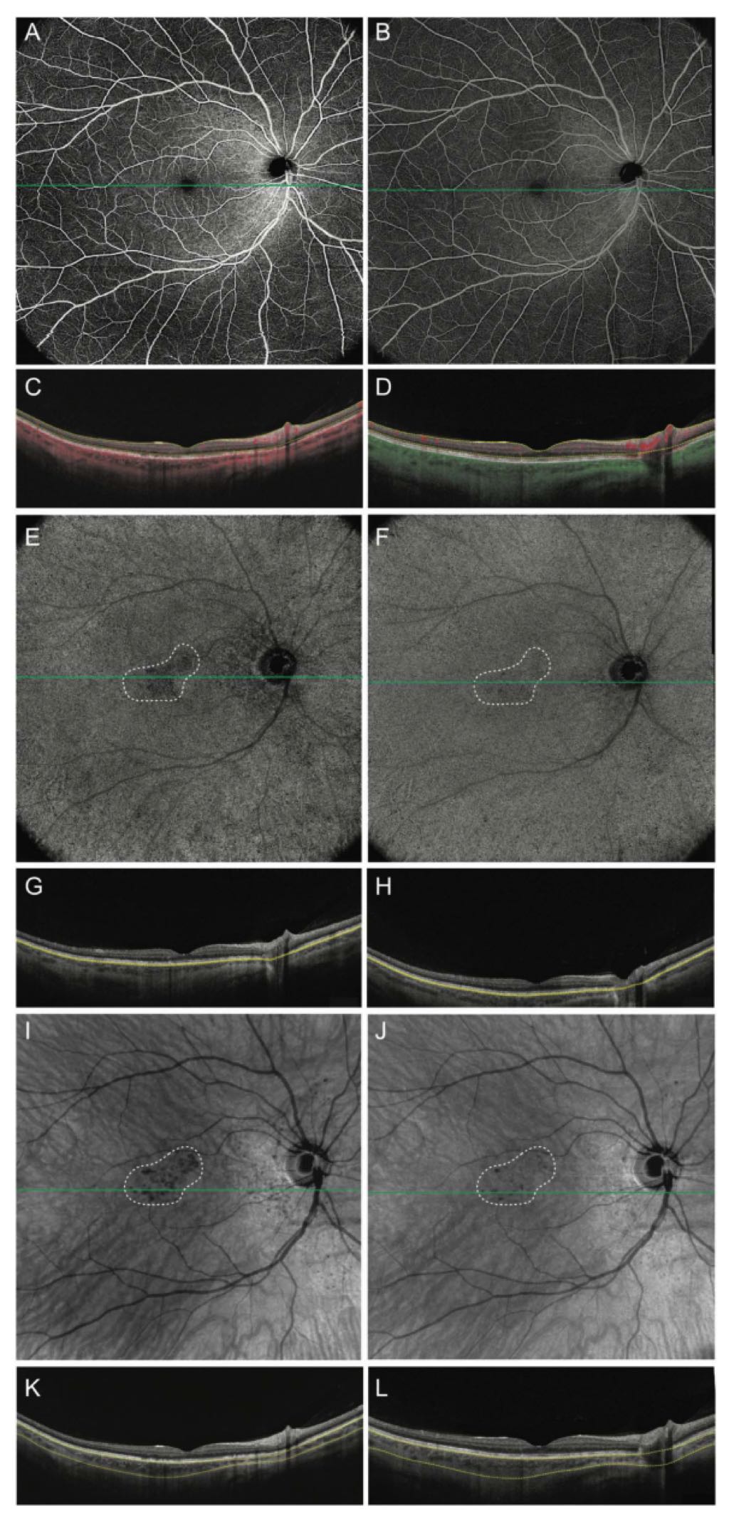
Wide-field SS-OCTA:
Revealed choroidal flow voids (subfoveal and peripapillary), consistent with choroidal infarction.
Retinal vasculature remained normally perfused.
Follow-up (6 months):
Visual acuity improved.
OCTA showed partial resolution of choroidal flow voids.
Infrared imaging revealed coalescing RPE changes.
🔬 Pathophysiological Insights
Choroidal vessels lack autoregulation, making them vulnerable to ischemia during hypertensive crises.
sFlt-1, a soluble VEGF receptor released during pregnancy, may contribute to endothelial dysfunction.
Choroidal infarcts can lead to RPE dysfunction and serous retinal detachments due to breakdown of the outer blood-retinal barrier.
🧪 Imaging Modality Significance
Wide-field SS-OCTA offers a noninvasive, high-resolution alternative to dye-based angiography.
Especially valuable in pregnant or breastfeeding patients where fluorescein or indocyanine green angiography may be contraindicated.
📝 Conclusion
HELLP syndrome can cause localized choroidal infarction detectable via SS-OCTA.
This imaging modality is crucial for diagnosing and monitoring retinal and choroidal vascular complications in pregnancy-related hypertensive disorders.
📸 Figure 1: Multimodal Imaging of Perifoveal Placoid Lesion
Panels A & B: Color fundus photographs show a polygonal placoid lesion with hyperpigmentation and Elschnig spots near the optic nerve.
Panel C: OCT reveals ellipsoid zone disruption and subretinal hyperreflective deposits.
Panel D: Infrared imaging highlights the hyperreflective placoid lesion.
Panels E & F: Follow-up OCT and infrared images show improvement—condensation of hyperreflective material into discrete deposits and partial resolution of the lesion.