Visual Loss in Geographic Atrophy

Visual Loss in Geographic Atrophy

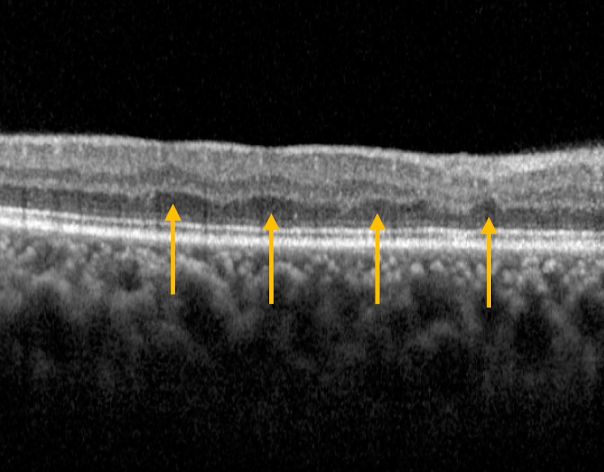
This study examines factors influencing visual loss in geographic atrophy (GA) related to age-related macular degeneration. Researchers analyzed clinical trials and observational studies to explore correlations between baseline visual acuity, lesion size, growth rate, foveal involvement, and focality with changes in visual acuity over two years. Key findings reveal faster GA lesion growth correlates with greater vision loss, particularly in eyes with subfoveal or unifocal lesions. Baseline measures, like visual acuity and lesion size, do not predict vision changes. Foveal involvement significantly accelerates vision loss. The study emphasizes identifying high-risk patients and highlights the need for interventions to slow GA progression.
AMD and Ultra-Wide Field Imaging

AMD and Ultra-Wide Field Imaging

This 5-year study used ultra-widefield imaging to track peripheral retinal changes in 265 eyes of 137 participants from the OPERA ancillary study of AREDS2. Researchers found that while AMD severity increased in the central retina, peripheral AMD features remained common and relatively stable. Interestingly, non-AMD peripheral changes like cobblestone degeneration became more frequent. Notably, the presence of significant peripheral findings did not appear to increase the risk of progression to advanced AMD. The study suggests AMD has pan-retinal involvement, but peripheral findings weren't predictive of advanced disease progression in this cohort.
Pupillometry and COVID

Pupillometry and COVID

This prospective study explored virtual reality-based infrared pupillometry (VIP) as a tool for detecting long COVID. Researchers recorded pupillary light responses in 185 participants using a VR headset and analyzed nine waveform features. They found that constriction time after the brightest light stimulus was significantly linked to long COVID. Machine learning models, particularly an LSTM model analyzing the full pupillary response, achieved high accuracy (up to 93.75%) in distinguishing long COVID from both post-COVID individuals and healthy controls. The study concludes that VIP shows potential as a non-invasive, affordable, and portable method for the objective detection and monitoring of long COVID, representing the first use of VR-based pupillometry for this purpose.
Recurrent RRD after Vaginal Versus Cesarean Delivery

Recurrent RRD after Vaginal Versus Cesarean Delivery

This retrospective study investigated retinal redetachment risk in women with a history of the condition, comparing vaginal and cesarean deliveries. Analyzing 66 eligible patients from an initial group of 967, the researchers found no statistically significant difference in redetachment rates: 5% after vaginal delivery versus 7.5% after cesarean. They conclude that cesarean delivery offers no ophthalmic benefit in preventing redetachment and doesn't recommend it for women with prior retinal detachment.
Omega-3 Fatty Acids and Age-related Macular Degeneration

Omega-3 Fatty Acids and Age-related Macular Degeneration

This study used both a prospective cohort study and Mendelian randomization (MR) to investigate whether omega-3 fatty acids protect against age-related macular degeneration (AMD). The cohort study, involving 258,350 participants from the UK Biobank, found that higher plasma levels of omega-3 and DHA were associated with a lower risk of developing AMD. MR analyses further suggested a causal link between a genetic predisposition to higher omega-3 and DHA levels and a reduced risk of both dry and wet AMD. The authors conclude that these findings support the need for clinical trials to assess the effectiveness of omega-3 fatty acids in preventing and treating AMD.
Retinal ischemic perivascular lesions (RIPLs) are linked to single subcortical infarction (SSI)

Retinal ischemic perivascular lesions (RIPLs) are linked to single subcortical infarction (SSI)

This research highlights the connection between retinal ischemic perivascular lesions (RIPLs), observed through noninvasive eye imaging (OCT/OCTA), and single subcortical infarction (SSI), a type of stroke. The study found that the presence of RIPLs is independently associated with SSI and correlates with a higher burden of cerebral small vessel disease in SSI patients, including increased white matter lesions and enlarged perivascular spaces in the brain. Additionally, eyes with RIPLs showed lower blood vessel density in a specific layer of the retina. These findings suggest that RIPLs could serve as a readily detectable marker of both retinal and cerebral ischemia, potentially aiding in the early identification of individuals at risk for or affected by small vessel disease in the brain.