Acute Myeloid Leukemia
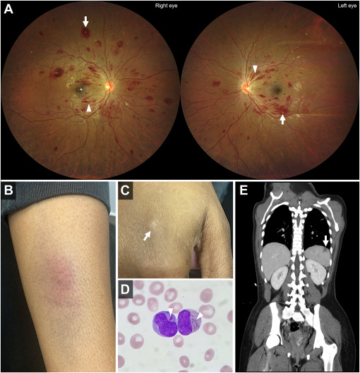
The fundus examination of a 22-year-old woman in the eye emergency department revealed bilateral white-centered hemorrhages, flame-shaped hemorrhages, and macular preretinal hemorrhage. The medical interview indicated recent weakness, dyspnea on exertion, and left hypochondrial pain. Physical examination showed skin bruises and subcutaneous nodules. Blood cell count indicated anemia, thrombocytopenia, and hyperleukocytosis. Blood smear revealed Auer rods. Computed tomography scan displayed a hypodense spleen lesion suggestive of splenic infarction. The diagnosis of acute myeloid leukemia was confirmed by flow cytometry on bone marrow aspiration. Urgent chemotherapy (idarubicin-cytarabine) was required.
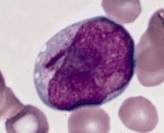
Auer rods, also known as Auer bodies, are crystalline cytoplasmic inclusion bodies found in myeloid blast cells during conditions such as acute myeloid leukemia, acute promyelocytic leukemia, high-grade myelodysplastic syndromes, and myeloproliferative disorders. These structures are composed of fused lysosomes rich in lysosomal enzymes and can take on various shapes like needles, commas, diamonds, rectangles, corkscrews, or granules. Although named after John Auer, they were first identified by Canadian physician Thomas McCrae in 1905, and both McCrae and Auer initially misinterpreted the cells containing these rods as lymphoblasts.
Reference : Sight Threat, Life Threat – Ophthalmology Retina