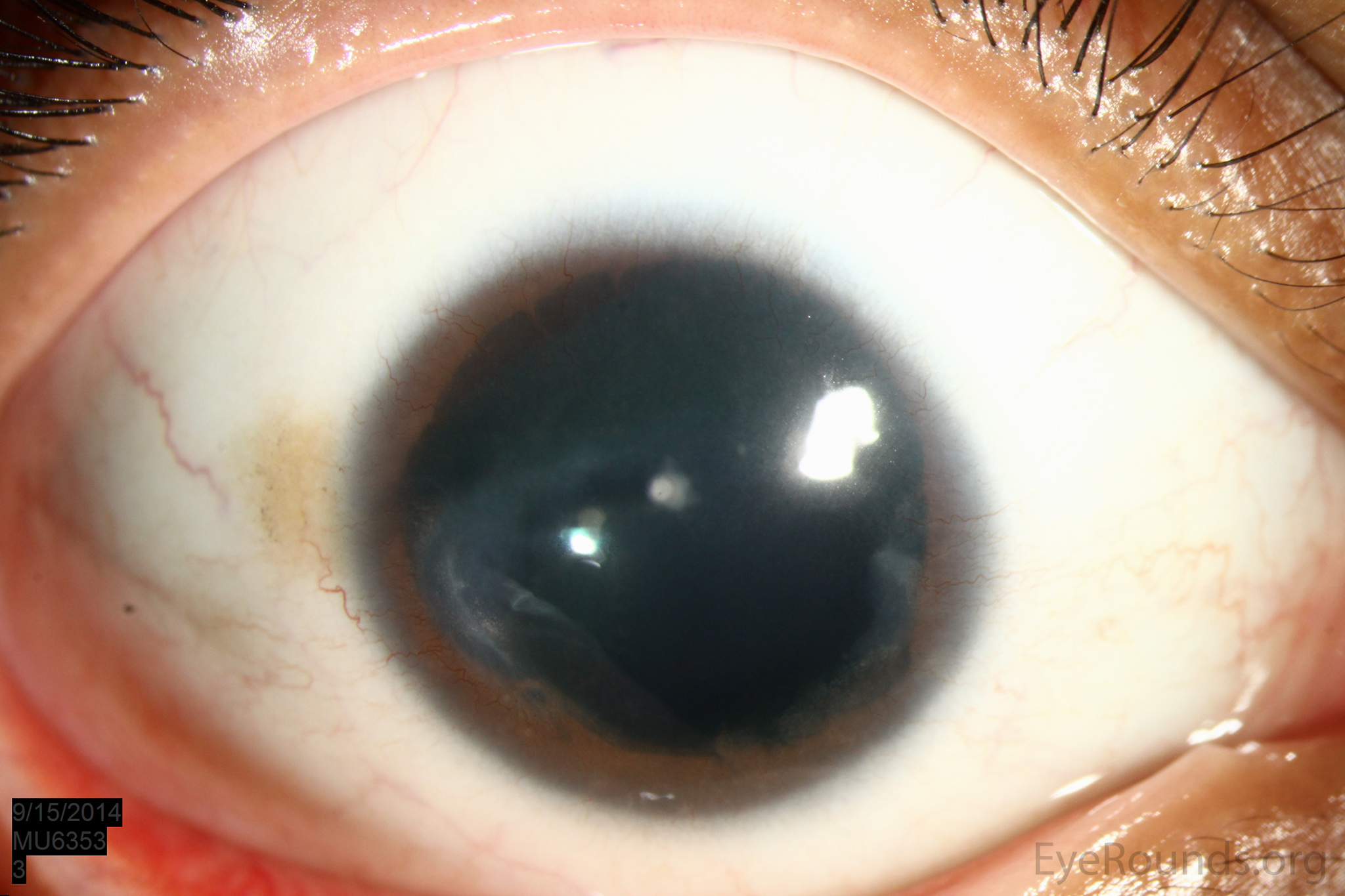
رشد طبیعی foveal در رحم در midgestation با جابجایی گریز از مرکز لایه های شبکیه داخلی (IRLs) از محل فووای اولیه اغاز می شود. تغییرات بیرونی شبکیه مانند افزایش تعداد سلول های مخروطی، کشیدگی سلولهای مخروطی و متراکم شدن آنها عمدتا پس از تولد رخ می دهد و تا سن ۱۳ سالگی ادامه می یابد. بلوغ فووا را می توان با استفاده از OCT ارزیابی کرد که در Foveal Pit به خوبی توسعه یافته، اکستروژن لایه های شبکیه داخلی،ضخیم شدن لایه هسته ای خارجی و کشیدگی بخش های خارجی فوتورسپتورها دیده میشوند. ناهنجاری های رشدی در درجات مختلف می تواند منجر به هیپوپلازی فوویال شود. هیپوپلازی فوویال به عنوان مثال در البینیسم، انیریدیا، نارسی، و هیپوپلازی عصب بینایی دیده میشود.
در مقاله جالبی که در مجله American Journal of Ophthalmology چاپ شده است با بررسی بیماران مبتلا به آنیریدیای مادرزادی به کمک تصویربرداری OCTA فرضیه جالبی در مورد تکامل Foveal Pit مطرح کرده است.
Optical Coherence Tomography Angiography Assessment in Congenital Aniridia
Am J Ophthalmol. ۲۰۲۳ Apr 13;253:44-48. doi: 10.1016/j.ajo.2023.04.004. Online ahead of print
ABSTRACT
PURPOSE: This study aims to characterize foveal vasculature assessed by optical coherence tomography angiography (OCT-A) in congenital aniridia which is hallmarked by foveal hypoplasia (FH).
DESIGN: Cross-sectional case-control analysis.
METHODS: At the National Referral Center for congenital aniridia, patients with confirmed PAX6-related aniridia and FH diagnosed on spectral-domain OCT (SD-OCT) with available OCT-A and matched control subjects were included. OCT-A was performed in patients with aniridia and control subjects. Foveal avascular zone (FAZ) and vessel density (VD) were collected. VD in the foveal and parafoveal areas at the level of the superficial and deep capillary plexi (SCP and DCP, respectively) were compared between the 2 groups. In patients with congenital aniridia, correlation between VD and the grading of FH was assessed.
RESULTS: Among 230 patients with confirmed PAX6-related aniridia, high-quality macular B-scans and OCT-A were available in 10 patients.
On the foveal area, mean VD was higher in aniridia patients (41.10%, n = 10) than in control subjects (22.65%, n = 10) at the level of the SCP and the DCP (P = .0020 and P = .0273, respectively).
On the parafoveal area, mean VD was lower in patients with aniridia (42.34%, n = 10) than in healthy subjects (49.24%, n = 10) at the level of both plexi (P = .0098 and P = .0371, respectively).
In patients with congenital aniridia, a positive correlation was found between the grading of FH and the foveal VD at the SCP (r = 0.77, P = .0106).
CONCLUSIONS: Vasculature is altered in PAX6-related congenital aniridia, higher in foveal and lower in parafoveal areas, especially when FH is severe, which is consistent with the concept that the absence of retinal blood vessels is essential for foveal pit development.
PMID:۳۷۰۵۹۳۱۶ | DOI:۱۰.۱۰۱۶/j.ajo.2023.04.004